Forum
Implantologicum

December 3, 2025
// 20 min

The Use of Tissue Level Design Implants in Immediate Protocols

Thomas
Gill
Michael
Myint
Shakeel
Shahdad

Abstract

The tissue level (TL) implant is considered a challenging implant for the esthetic zone as the perceived potential esthetic risk of exposure of the machined collar or metal shine-through commonly drives clinicians to select bone level (BL) design implants, especially for immediate implant placement (IIP) protocols. Despite using BL design implants, studies investigating the outcomes of IIP have reported a high frequency of facial mucosal recession. In our experience, TL design implants provide comparable outcomes and seem to have been unfairly implicated for esthetic failures. 

This article provides a narrative review of the evolution of TL implants and outlines the benefits of this design for IIP. Additionally, clinical cases are utilized to highlight the effectiveness of TL implants for IIP in the esthetic zone. 

Based on the current evidence, there is a paucity of studies assessing the stability of clinical outcomes for IIP with TL implants in the esthetic zone. The standard indications and contra-indications apply regardless of implant type, and the optimal restoratively driven three-dimensional position of the implant should not be compromised.

Introduction

The tissue level (TL) implant has been at the forefront of innovation for more than four decades and is one of the oldest and most well-evidenced implants used in modern dentistry. At a time when Brånemark implants followed a fully submerged healing protocol, the one-piece TL (ITI) implants were novel, pushing the boundaries to pioneer transmucosal healing (Brånemark et al. 1969; Buser et al. 1990; Buser et al. 1997; Andersen et al. 2002).

As time progressed, surgical protocols were refined to allow for earlier placement and predictable loading protocols. Over the last two decades, the demands and benefits of immediate implant placement (IIP) have continued to increase from a patient and clinician perspective (den Hartog et al. 2008; Slagter et al. 2014; Huynh-Ba et al. 2018; Morton et al., 2018; Morton et al., 2023). IIP is defined as the placement of a dental implant into a fresh socket on the same day as tooth extraction (Gallucci et al. 2018).

Many readers might find it surprising that several early studies on IIP in the esthetic zone utilized TL implants as this was what was largely available at the time (Lang et al. 1994; Bragger et al. 1996; Andersen et al. 2002; Bianchi & Sanfilippo 2004). In the 1990s, these implants were parallel-sided with passive thread designs and, with developing IIP protocols, were considered technically demanding to place and lacked sufficient primary stability for IIP. From 2000 onwards, bone level (BL) design implants gained preference and have been predominantly used in research studies, with the introduction of implant geometries that offer relatively predictable high primary stability at implant insertion (Cochran et al. 2016; Dard et al. 2016). Until recently, implant geometries that aid immediate protocols have been largely absent from manufacturer portfolios for TL implants, and therefore, modern studies have rarely reported outcomes of TL implants (Wittneben et al. 2023). However, the authors believe TL implants can be successfully and predictably utilized for IIP in the anterior esthetic zone and achieve stable long-term esthetic outcomes. 

It is to be reiterated that replacing a single tooth in the anterior maxilla (#15 to #25) with an IIP protocol is a complex procedure. The rules for placing TL immediate implants in the anterior maxillary esthetic zone are similar to those for BL design implants. IIP may be considered the treatment of choice when ideal conditions are present (Wittneben et al. 2023), such as:

  • Healthy adjacent teeth
  • Thick gingival phenotype
  • Intact facial bone >1 mm in thickness
  • No acute infection or chronic fistula
  • Ability to place the implant in the correct three-dimensional position for a screw-retained restoration
  • Ability to achieve primary stability of the implant, which is a fundamental prerequisite if an immediate restoration is expected.
 

This article aims to discuss the tissue level implant’s potential advantages, address concerns about its use, and highlight the esthetic outcomes achievable when immediately placing TL implants in the esthetic zone.

The tissue level implant

The tissue level (TL) implant is characterized by two main sections: 

  1. a rough implant body, and 
  2. a smooth machined collar at the coronal end.
 

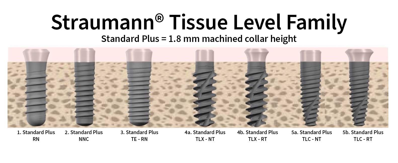
The TL design has a distinct rough-smooth junction (Fig. 1), with the rough surface aimed to promote “osseointegration” and the machined collar “mucointegration” for a stable transmucosal peri-implant soft tissue (Herman et al. 2000; Hermann et al. 2011). The design allows soft tissue morphogenesis to commence immediately after placement, which then remains undisturbed during healing and shifts the implant-abutment junction vertically away from the crestal bone (Hermann et al. 2001; Hanggi et al., 2005; Sasada & Cochran 2017). Most commonly, the machined coronal component measures 1.8 – 2.8 mm for the establishment of “the biologic width” (De Sanctis et al. 2010; Parvini et al. 2023). Given the pronounced gingival scalloping around natural teeth, 1.8 mm collar height is the preferred choice for the esthetic zone. Several manufacturers produce TL implants, among them: Thommen, Sweden & Martina, Biohorizon Camlog, Osstem, Institut Straumann AG, although the authors’ experience has mostly been with the latter.

The macro-geometry of TL implants (Institut Straumann AG, Switzerland) can be broadly separated into four designs (Fig. 1): 

  1. Parallel-sided
  2. Coronally tapered
  3. Fully tapered
  4. Apically tapered

Parallel-sided, passive threaded implants, such as the conventional Straumann® Tissue-level SP implants, were the first TL implant geometry used for IIP in the esthetic zone, with excellent survival rates (Lang et al.1994; Gher et al. 1994; Lang et al. 1994; Bragger et al. 1996).  

As IIP protocols developed, high primary stability for immediate loading became increasingly desirable (Javed et al. 2010). In the anterior maxilla, the wider root form at its coronal aspect commonly makes it difficult to achieve an optimal restoratively driven implant position with adequate primary stability using the parallel-sided passive thread design. This led to the early development of coronally tapering TL implants (Straumann® TE implant), mirroring the root form of the extracted tooth to increase the engagement of socket walls and increase mechanical stability (Akkocaoglu et al. 2005; Linares et al. 2011). At 5 years, the survival rate of these coronally tapering TL implants (97.7%) was comparable to other implant types (Roccuzzo et al. 2013; Wilson Jr 2013). However, reporting on esthetic outcomes was ignored in these earlier studies. Largely, coronally tapered implants have fallen out of favor due to a more profound understanding of the need for space between the implant and the buccal bone (Caneva et al. 2010).

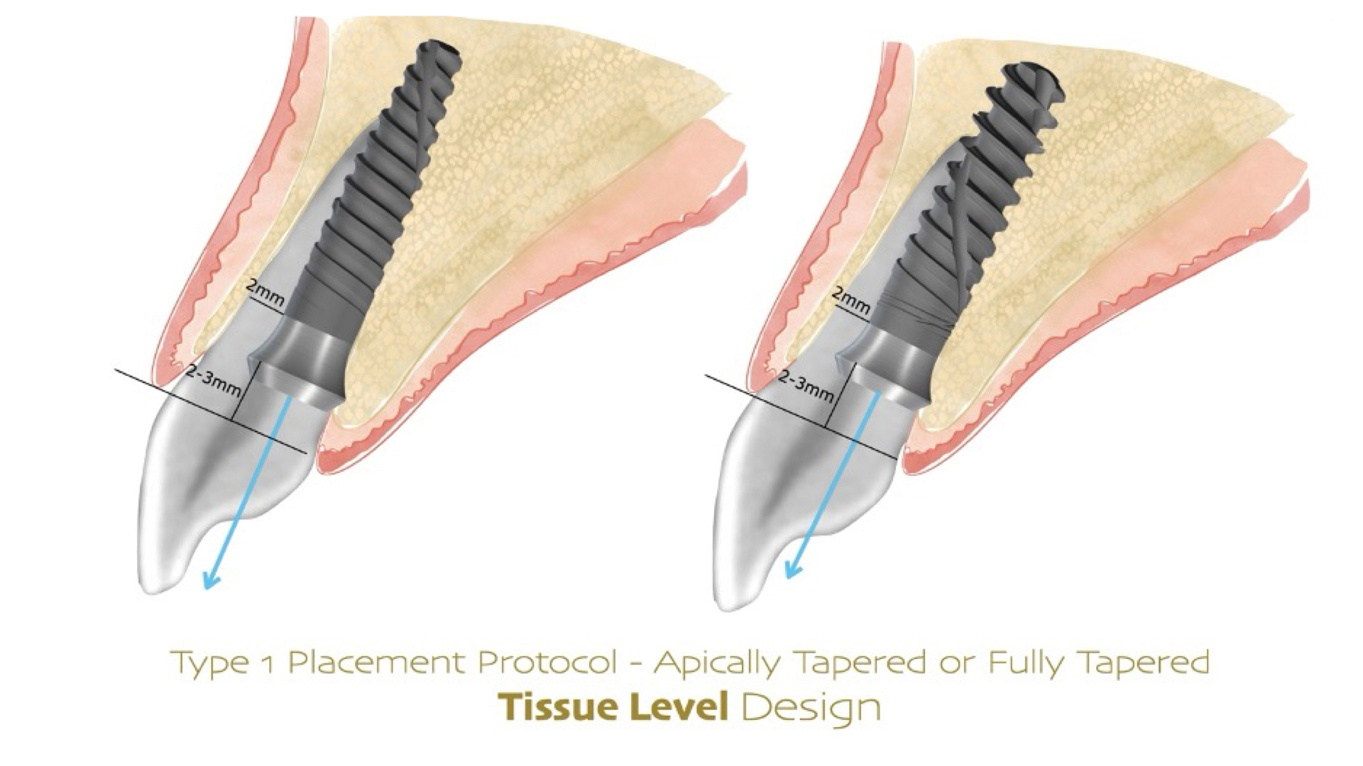
As implant geometries evolved, it became clear that tapering implants offered higher primary stability for IIP. The tapered apex of the implant ensures better primary stability by reducing the drilling diameter and increasing the compression of the cancellous bone (Ellis et al. 2020). The survival rate of immediately placed tapered implants is comparable to that of conventional placement protocols (Schiegnitz et al. 2024). Recently, fully tapered and apically tapered TL implants have been developed that achieve significantly higher primary stability (El Chaar et al. 2021, Gill et al. 2024) and are today preferred for IIP cases. TLX and TLC implants differ in their geometry (Fig. 1). The TLX has a tapered core with self-cutting, deep threads, whereas the TLC is self-tapping, apically tapered and cylindrical in its coronal aspect. In the anterior maxilla where the preparation is positioned palatally and the implant is easily displaced buccally upon insertion, the TLC has become generally our preferred geometry.

Advantages of tissue level design implants

Peri-implant soft tissue attachment or “mucointegration” has been widely characterized and serves to establish a soft tissue barrier that maintains and protects marginal bone levels (Berglundh & Lindhe 1996; Salvi et al. 2015; Oskarsson et al. 2018; Bressan et al. 2023) and acts as a protective factor against peri-implant diseases (Tavelli et al. 2021). The repeated dis- and reconnections of the abutment in BL design implants has been demonstrated to result in a change in soft tissue architecture with a more apically positioned zone of connective tissue and additional marginal bone resorption (Berglundh & Lindhe 1996; Abrahamsson et al. 1997; Alves et al. 2005). Recently, this has led to the “one-time abutment” concept – a proposal to place a definitive abutment at implant placement which is not subsequently removed in order to preserve soft tissue attachment (Molina et al. 2017; Rathi et al. 2022; Calatrava et al. 2024; Sanz-Sanchez et al. 2024). This essentially attempts to convert a BL implant into a TL design, but with a horizontal platform shift.

The marginal bone loss observed around BL implants relates to the presence of connective-tissue inflammatory infiltrate due to the microgap at the implant-abutment junction relative to the crestal bone (Maeda et al. 2007; Liu & Wang 2017). Horizontal platform switching for BL implants, where a smaller diameter abutment is placed on a wider implant platform, demonstrated a reduction in marginal bone loss compared to platform-matching abutments (Lazzara & Porter 2006; Canullo et al. 2009; Canullo et al. 2010; Atieh et al. 2010; Strietzel et al. 2015; Oskarsson et al. 2018; Tomar et al. 2023). It is hypothesized that horizontal platform switching moves the microgap at the implant-abutment junction inwards away from the crestal bone (Lazzara & Porter 2006). 

On the contrary, the microgap in the TL implant is moved vertically away by typically 1.8 – 2.8 mm from the crestal bone and does not have an equivalent associated inflammatory infiltrate juxtaposed to the bone crest, thereby demonstrating less marginal bone loss (Broggini et al. 2003). Therefore, the relocation of this microgap vertically away, a “vertical platform shift”, from the crestal bone with the TL design contributes to the formation of robust soft tissues and preservation of crestal bone levels (Hermann et al. 2001; Hanggi et al. 2005; Sasada & Cochran 2017; Kim et al. 2018; Lago et al. 2018).

The TL design benefits from simultaneous bone and soft tissue healing, allowing for simultaneous osseo- and mucointegration, which is left undisturbed during the prosthetic phase of treatment. This leads to robust soft tissue seal and stable marginal bone levels, which may account for reduced risk of future peri-implant disease (Hermann et al. 2001; Derks et al. 2016).

For bone grafting, the tulip (divergent) shape of the TL implant provides additional benefits. Guided bone regeneration with TL implants resulted in thicker facial bone at the rough-smooth transition than at the shoulder of BL implants (Shahdad et al. 2013; Buser et al. 2013; Chappuis et al. 2016). We attribute this to the flared machined section of the TL implant, which provides space and protects the particulate bone used for bone regeneration (Shahdad et al. 2013; Quah et al. 2024). Furthermore, a third of TL implants exhibited bone vertical to the SLA-machined collar junction when the implant was placed >2 mm palatally to the adjacent teeth with facial bone >1 mm (Shahdad et al. 2023). This is a significant finding since it is assumed that the machined collar should have no bone around it.

In summary, the TL design offers the following advantages, that may be leveraged to optimize long-term outcomes in the esthetic zone: 

  1. Simultaneous biologic width establishment
  2. Vertically off-set microgap which moves the implant-abutment interface away from the crestal bone
  3. The soft tissues established during healing are not severed during abutment disconnection and reconnection
  4. Guided bone regeneration for stable hard and soft tissue peri-implant architectures supported.

Caution with immediate implant placement

Immediate implants are associated with greater variability in esthetic outcomes with a higher frequency of facial mucosal recession of >1 mm compared to early implant placement (Chen and Buser 2014; Hamilton et al. 2023). Careful assessment and analysis are crucial for appropriately determining any treatment plan, especially when proposing IIP treatment in the esthetic zone (Wittneben et al. 2023). An ethos of “minimizing morbidity without compromising long-term outcomes” should be prioritized. 

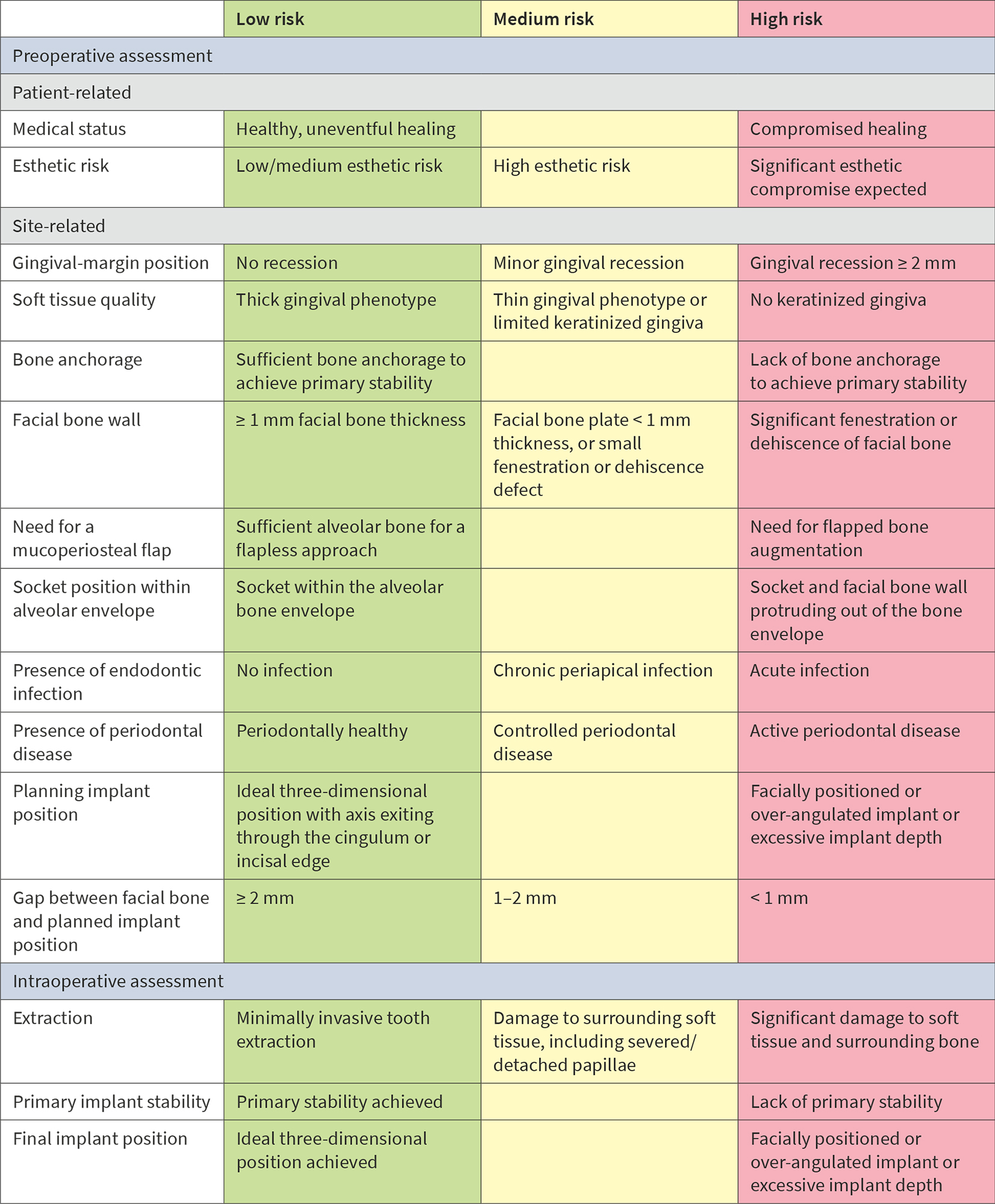
Patient and site-related factors must be considered for treatment to achieve predictable long-term functional and esthetic outcomes. The patient could be assessed using the Esthetic Risk Assessment (ERA) and risk assessment for IIP in single-tooth sites (Table 1) (Hamilton et al. 2023; Lambert et al. 2023) to determine the patient and site-specific risk factors for IIP. If the criteria for IIP protocol are not met, alternative placement protocols must be considered.

Optimizing esthetics for implant restorations in the anterior maxilla has been well studied, and implants should be restoratively driven irrespective of the timing of implant placement. It is critical to achieve an optimal, prosthetically-driven, three-dimensional implant position (Buser et al. 2004; Buser et al. 2017). In the anterior maxilla, midfacial recession has been reported for immediately placed BL implants in the presence of a thin (<1 mm) or missing facial bone plate (Chen et al. 2007; Seyssens et al. 2020). Further, clinical studies using CBCT scans to examine the facial bone on immediately placed implants have shown a significant relationship between recessions of the midfacial mucosa and a lack of visible facial bone on the CBCT images (Januário et al. 2011; Miyamoto & Obama 2011; Benic et al. 2012). The risks can be similar for TL implants, but the authors have experienced minimal risk of increased mucosal recession after placing TL implants in patients with <1 mm facial bone (Fig. 2). Although not currently supported by evidence, we attribute this to the simultaneous osseo- and mucosal integration that allows for better soft and hard tissue stability. Nevertheless, in some cases, loss of alveolar convexity has been noted, and the CBCT scans have confirmed the loss of alveolar convexity (Fig. 3).

After minimally traumatic extraction and preservation of facial bone plate, an immediate TL implant (NNC design) was placed (Type 1B) with simultaneous bone grafting in a 71-year-old male. The definitive restoration was placed 6 weeks later (Fig. 2c). For up to four years, the peri-implant tissues were stable with minimal recession, whilst there was a noticeable recession in adjoining natural teeth (Fig. 2d). There was minimal loss of alveolar contour despite <1 mm facial bone wall thickness (Fig. 2e). What is interesting to note here is that the bone is detectable coronal to the rough-smooth transition (Fig. 2f), which is not uncommon in our experience. Although histologically, this bone will not be “osseointegrated” with the machined collar, this thick, stable architecture achieved by the TL likely contributes to the long-term stable esthetics seen in our experience with TL implants.

In Figs 3a – c, a 75-year-old male requiring replacement of UL2 also has <1 mm facial bone thickness and pre-existing recession and a peri-apical lesion. Similar to the previous case, a Type 1B protocol with simultaneous GBR was chosen (Fig. 3d) (Gallucci et al. 2018). Loss of alveolar convexity was noted within weeks of provisionalization, even though the submarginal emergence of the crown was optimally shaped (Figs 3e – g). Nevertheless, the 1- and 2-year reviews show an excellent esthetic outcome (Figs 3h – k), which compares well with the pre-operative situation.

These two examples reiterate that outcomes with Type 1 protocols, especially the response of bundle bone resorption and its impact on bone graft particles, can be unpredictable, even with TL implants.

Interestingly, pre-clinical studies have consistently demonstrated that irrespective of implant type or dimension, resorption of bone after tooth extraction occurs (Araujo & Lindhe 2005; Caneva et al. 2010; Linares et al. 2011). Caneva and coworkers highlighted the need for sufficient space between the implant and the buccal bone. They studied the influence of the distance of the implant surface to the buccal bone for IIP by comparing 3.3-mm and 5-mm diameter implants. Marginal bone loss in wider diameter implants was more marked compared to the narrow diameter implants. This risk is heightened in thin phenotypes, so coronally tapered implants such as the Straumann TE are now not commonly used for IIP. Therefore, IIP in sites with a facial bone thickness of <1 mm should be considered a risk for loss of alveolar root convexity and midfacial recession. In such cases, if IIP is preferred, soft tissue augmentation at the time of placement should be considered (Seyssens et al. 2021).

One of the main concerns regarding TL in the esthetic zone is the risk of shine-through or exposure of the machined collar. In our experience, these complications result solely from surgical error and due to facial malpositioning of the implant (Chen et al. 2007; Chen et al. 2009), resulting in recession and esthetic failure (Chen & Buser 2009; Cosyn et al. 2019; Evans & Chen 2008). Most importantly, these complications arise irrespective of the chosen implant design.

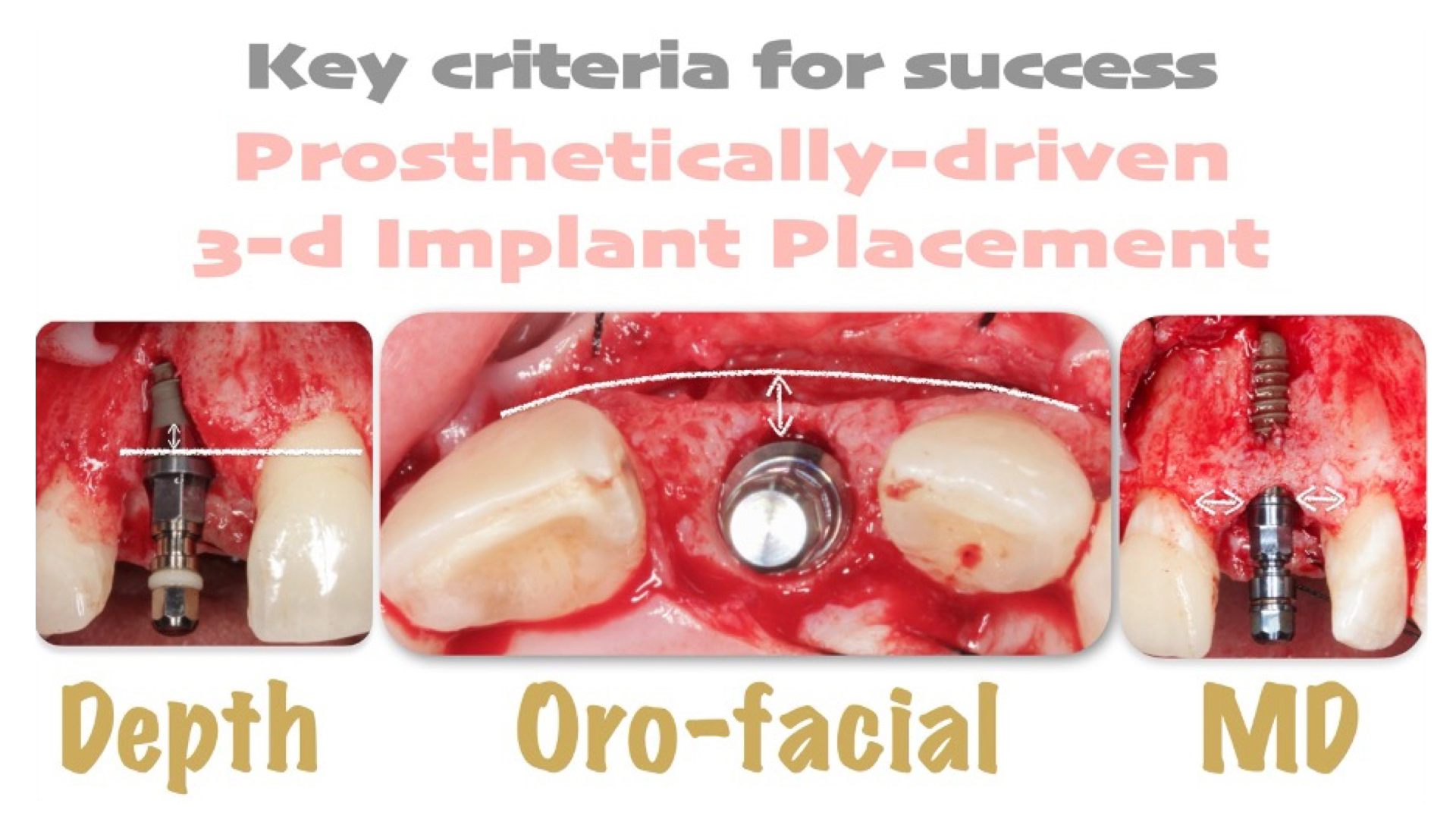
Tissue level implants in the Type 1 immediate protocol should be placed, adhering to the principles of 3-dimensional implant placement (Figs 4 – 5):

Apico-coronal position: not more than 2-3 mm apical to the proposed mucosal margin,

  1. Oro-facial position: ideally screw-retained with screw access through the cingulum. A minimum of 2-mm horizontal gap should be maintained between implant and internal socket wall at the junction of the rough surface,
  2. Mesio-distal position: >1.5 mm distance from implant shoulder to adjacent teeth.

The “tulip” shape of the machined collar needs to be accounted for in the osteotomy preparation with sufficient profiling. Similarly, the implant should be positioned towards the palatal wall for a screw-retained restoration, with the gap located buccally. In such cases, clinicians should be cognisant of the implant being inadvertently displaced facially during insertion (by the path of least resistance). This is most commonly encountered with implants with a more aggressive thread design.

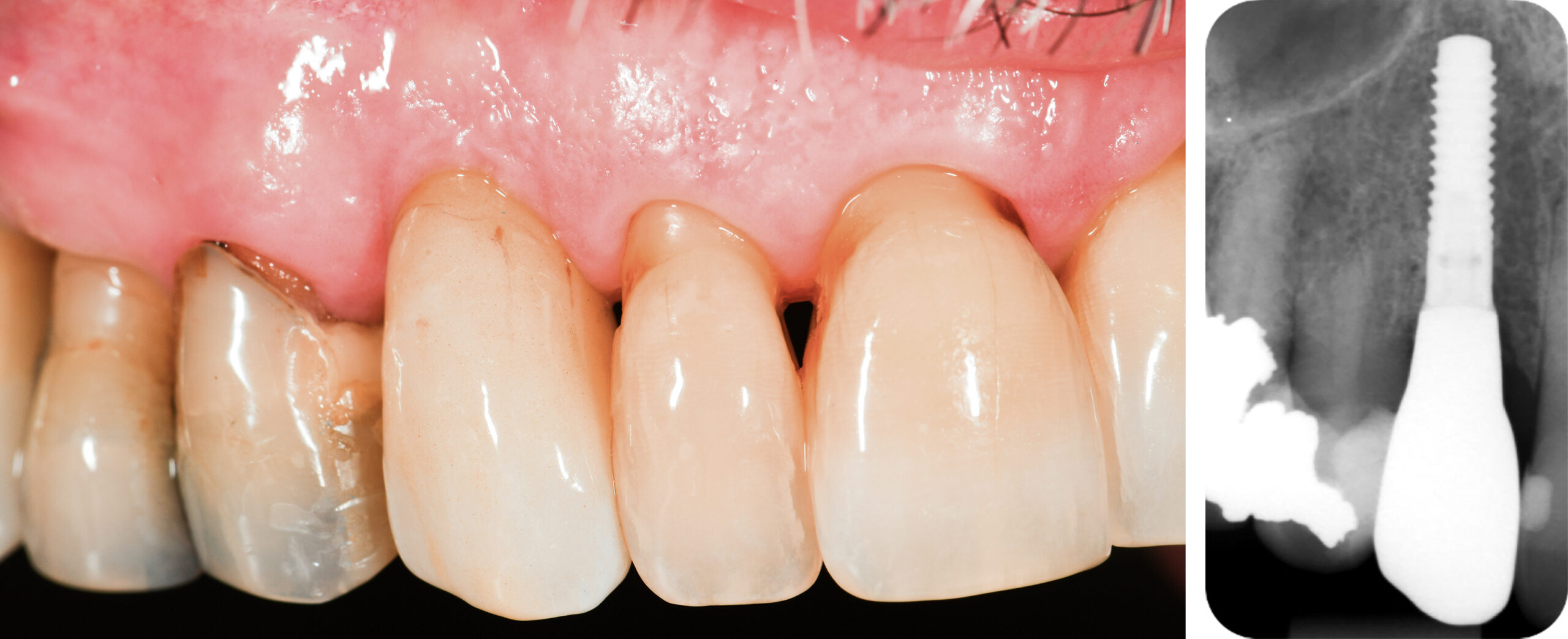
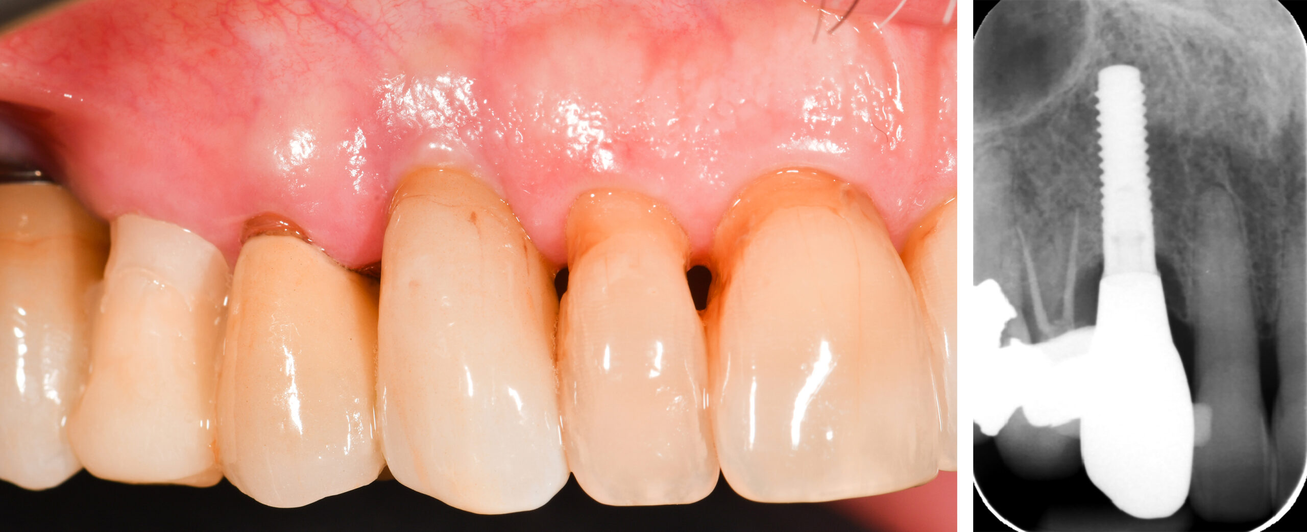
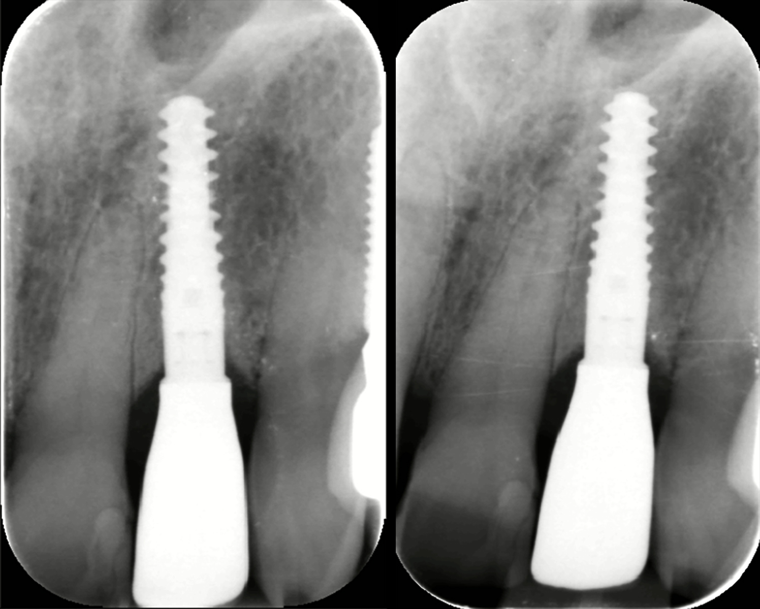
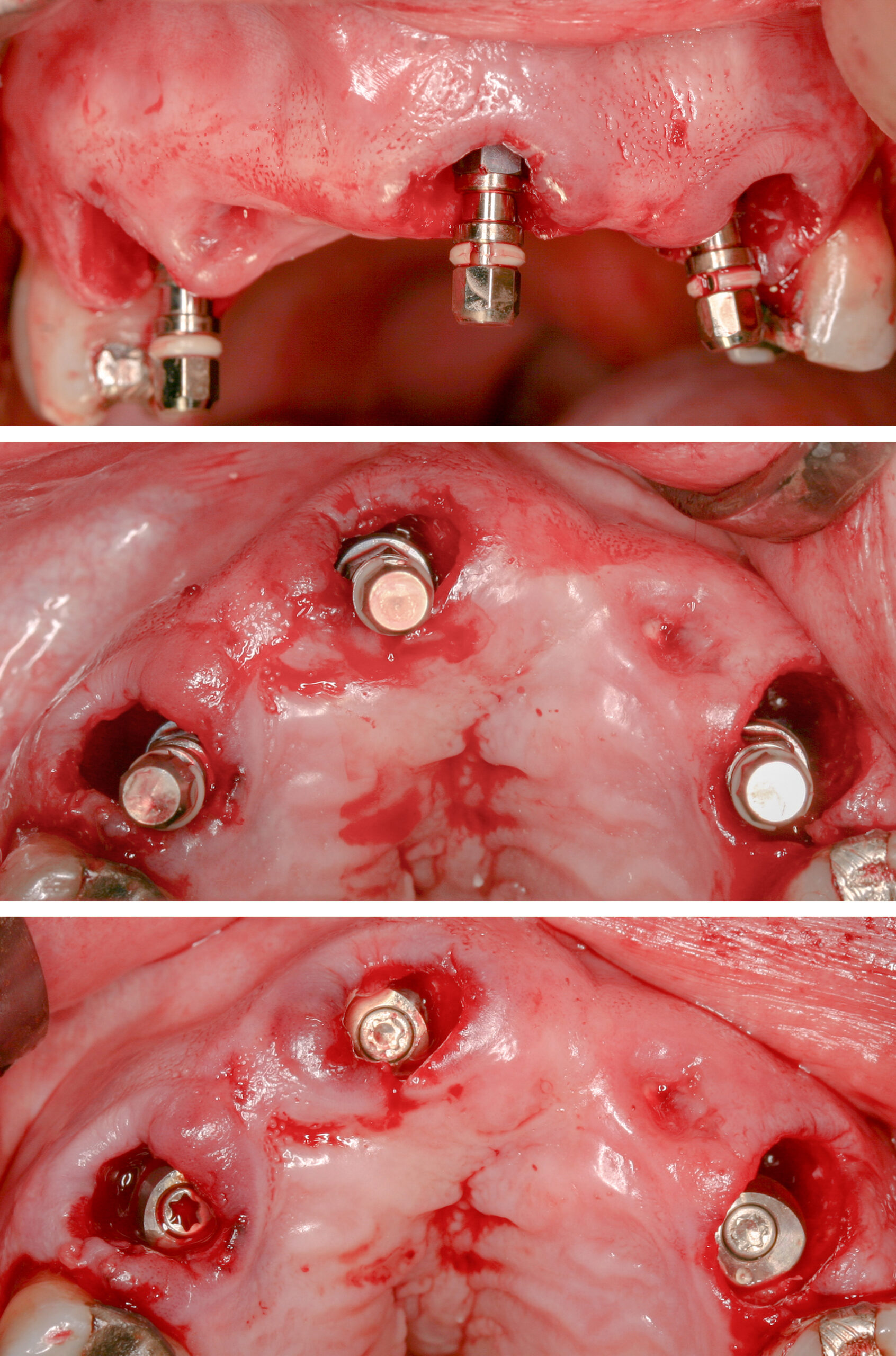
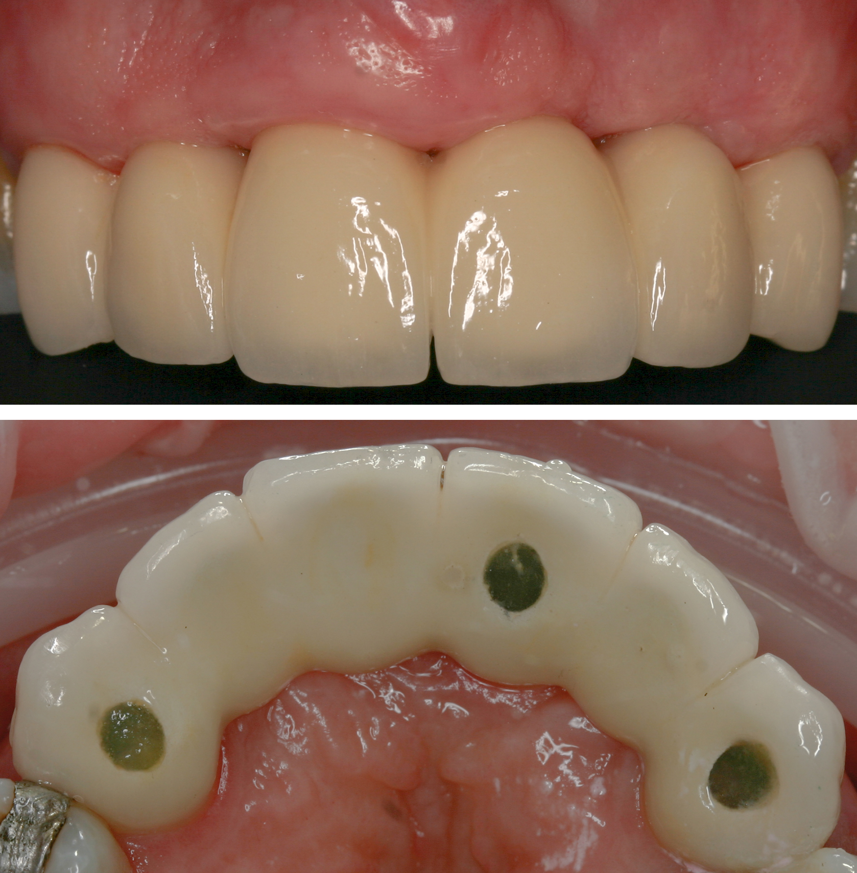
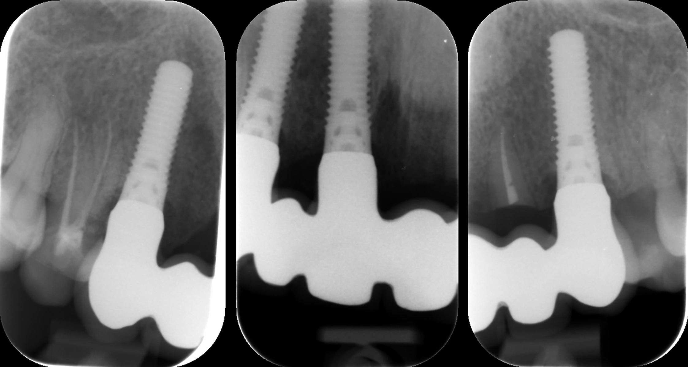
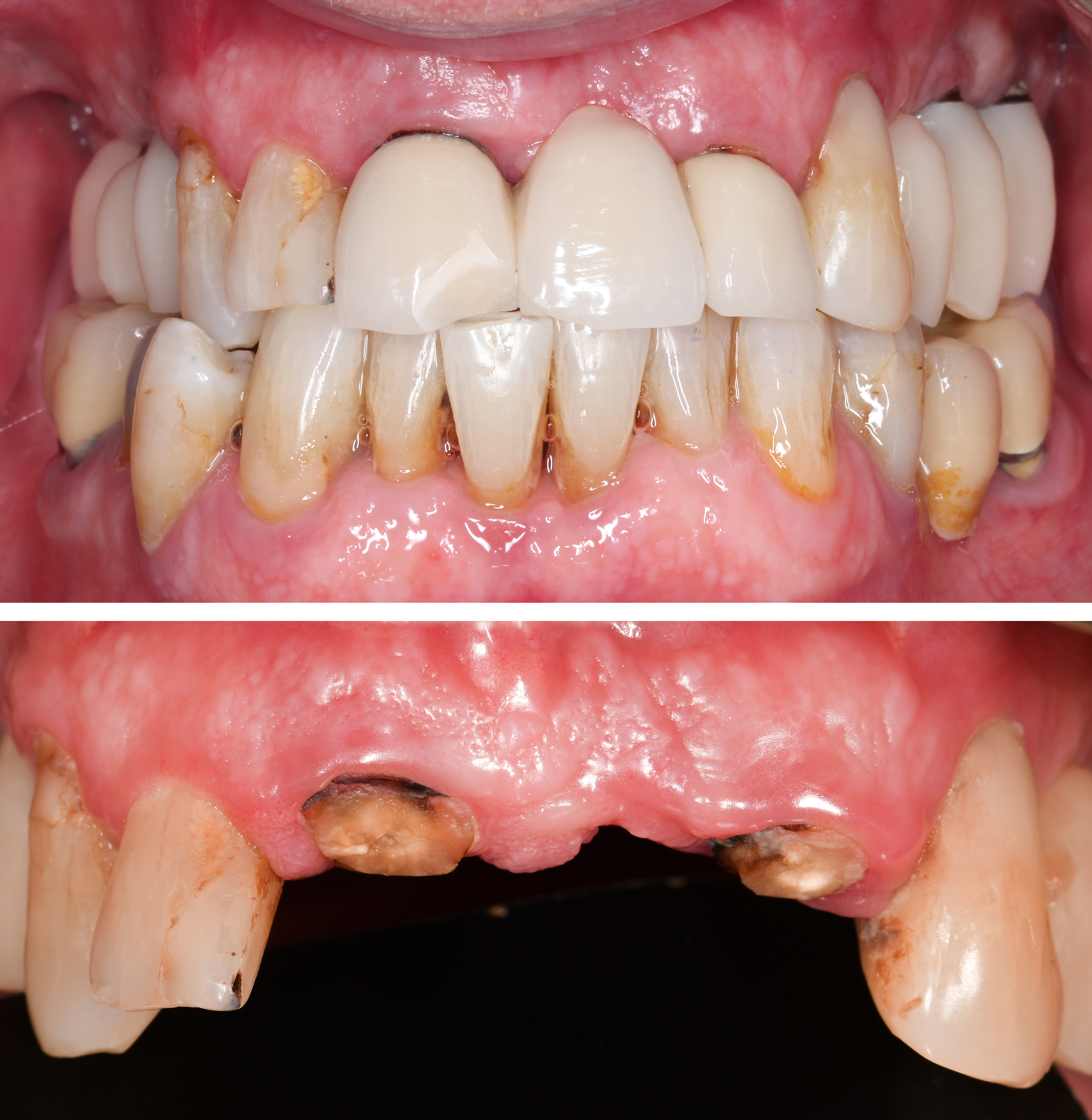
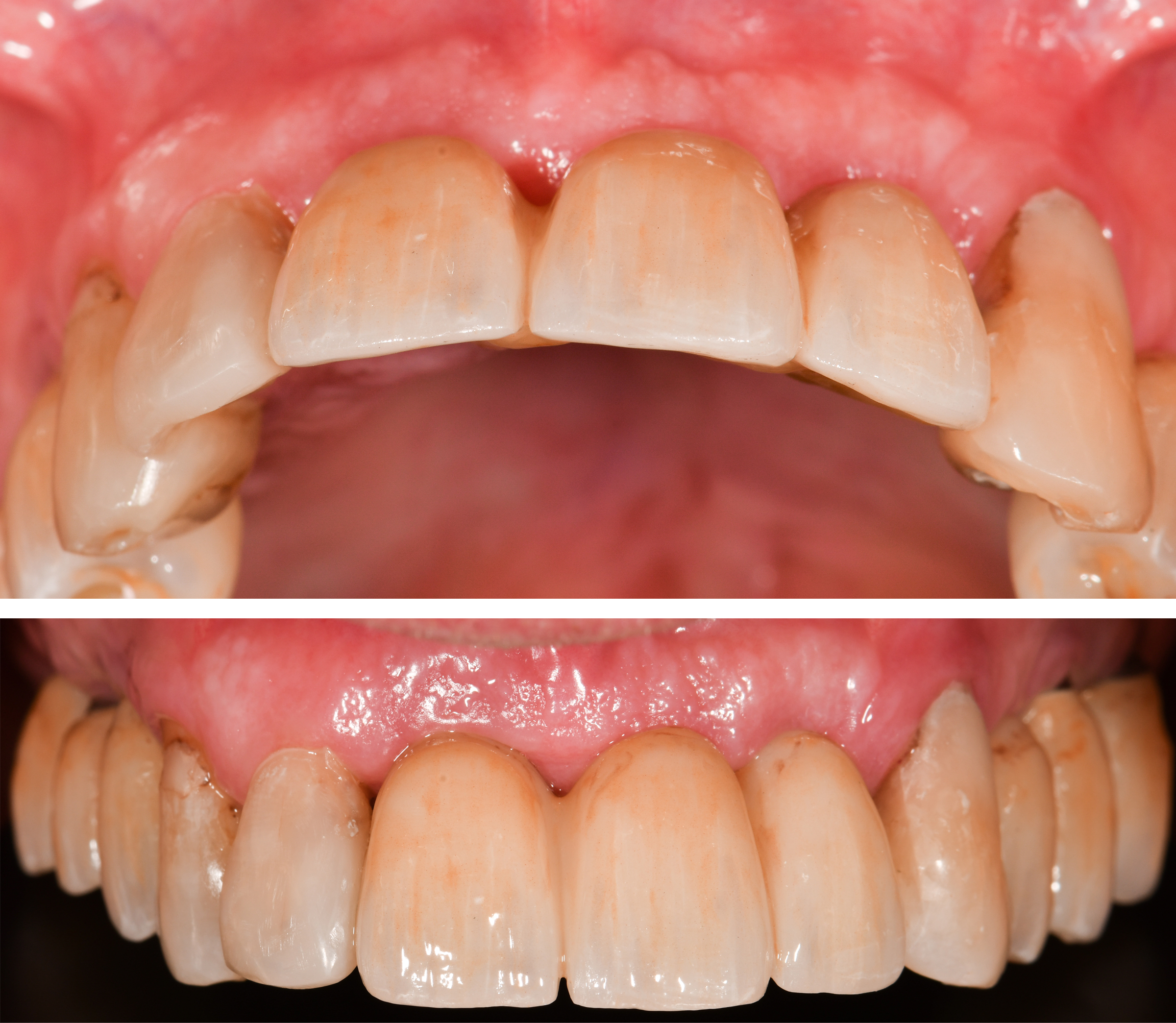
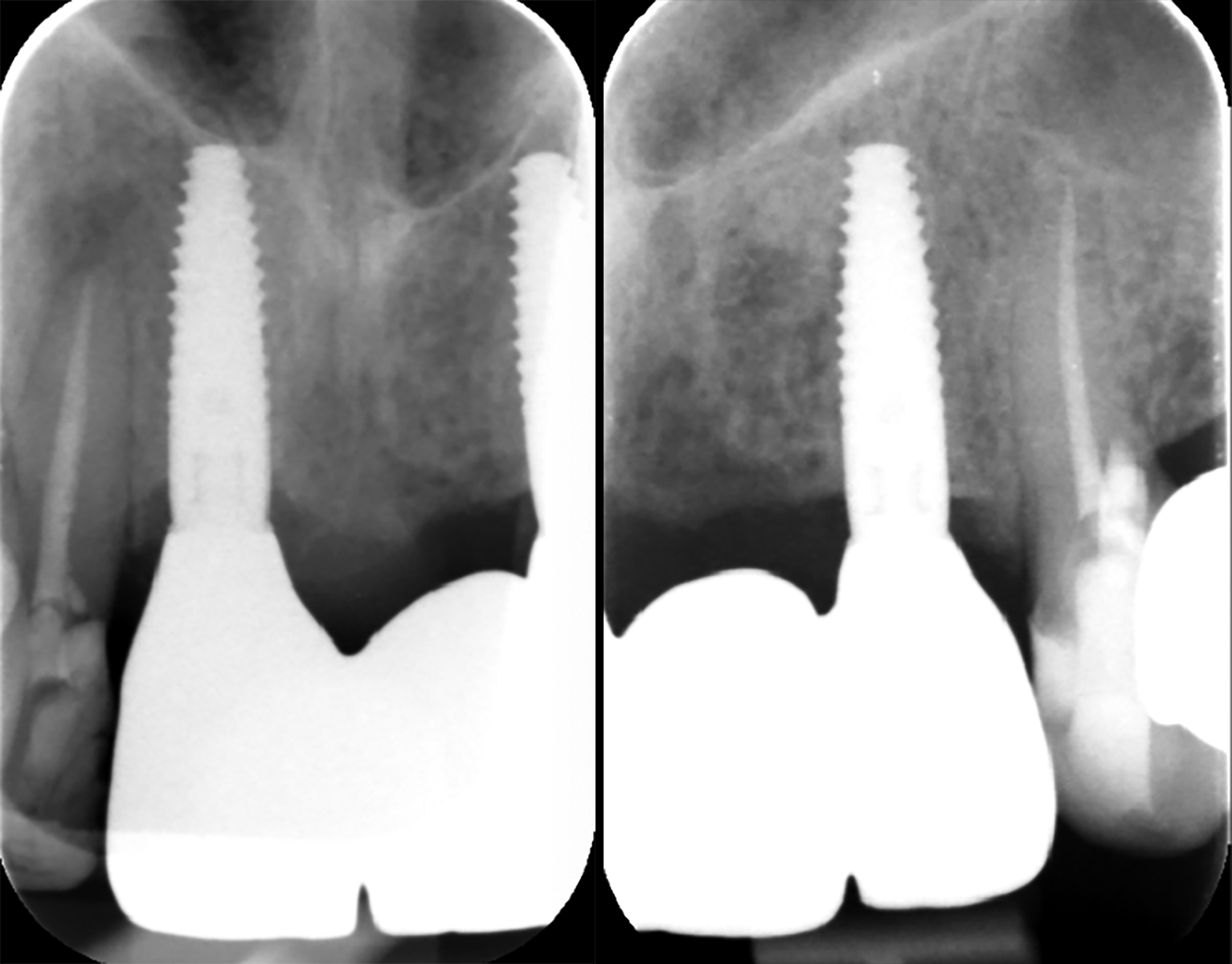
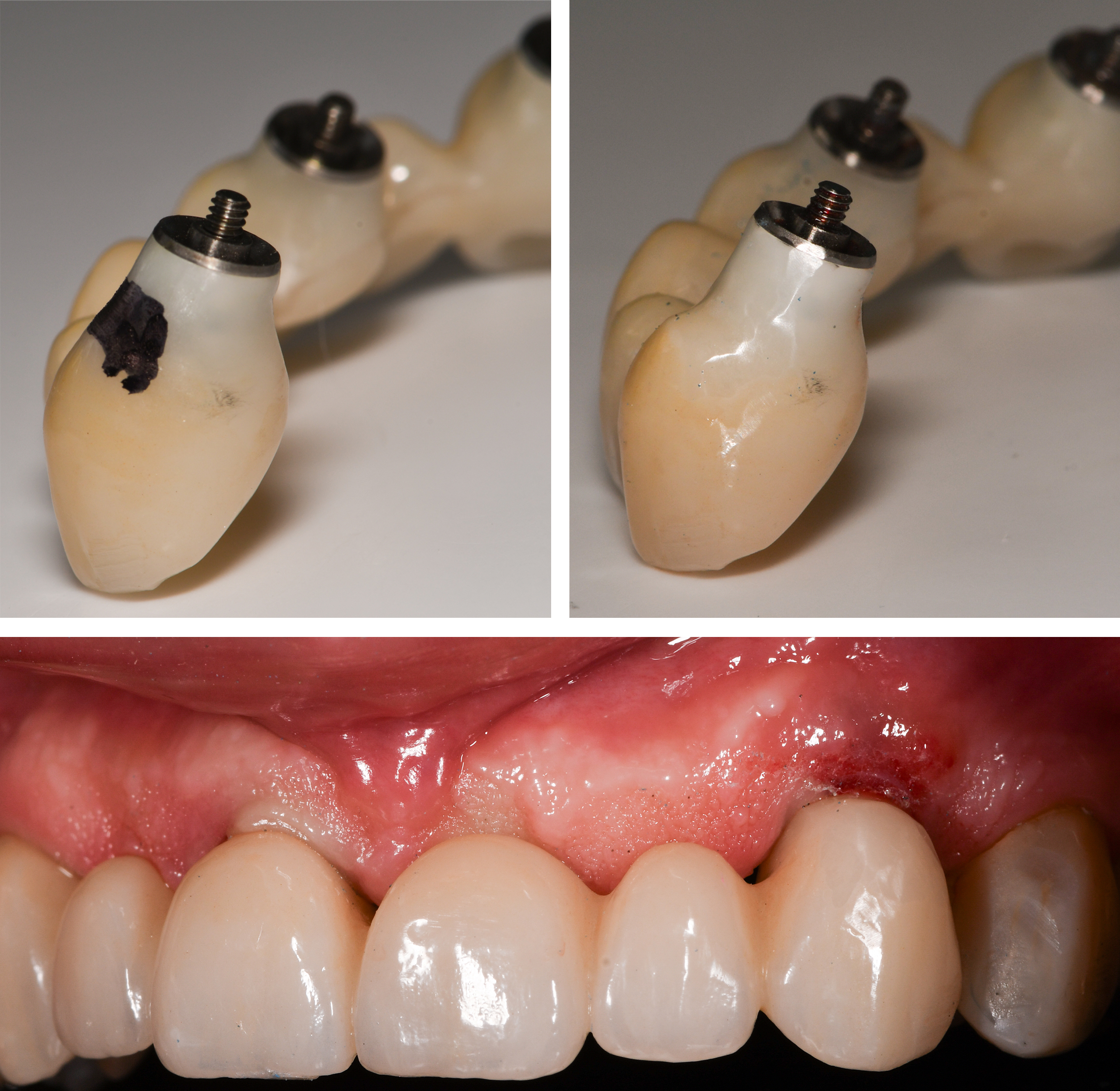
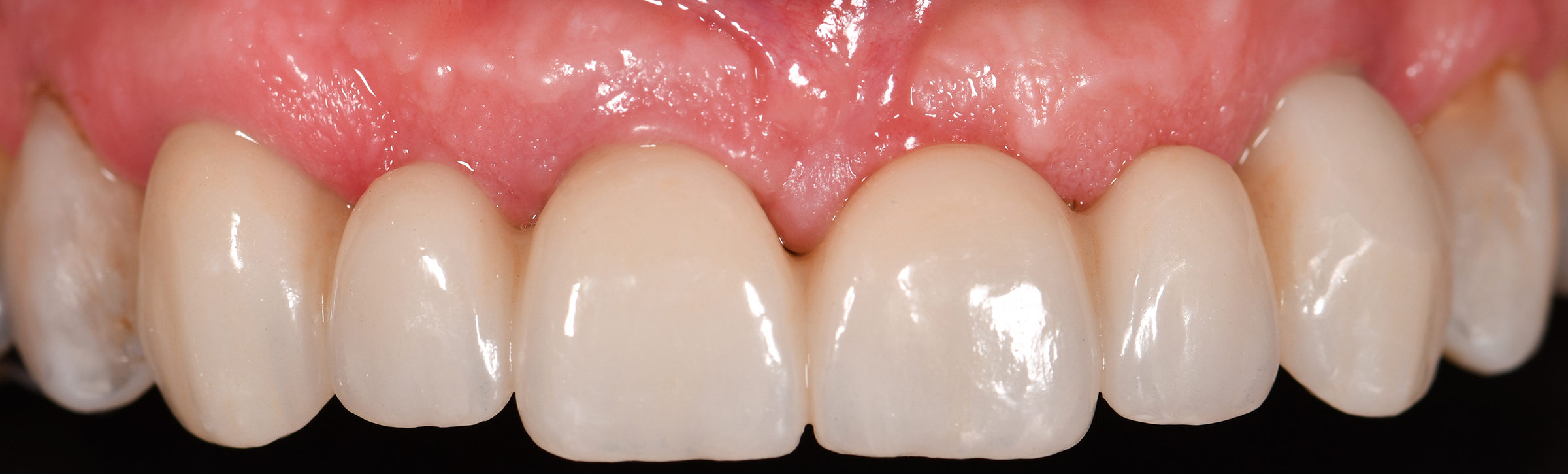
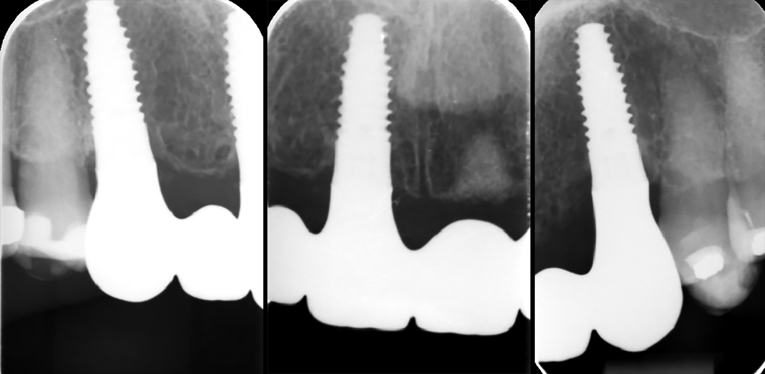
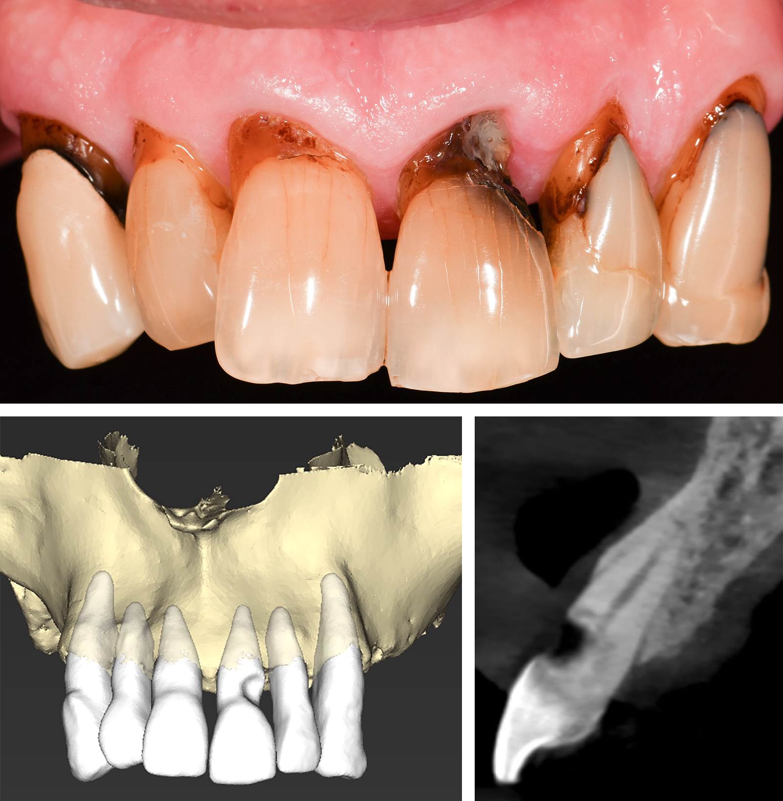
The clinical case in Fig. 6 pertinently highlights the importance of orofacial implant position and its impact on the labial bone. In 2009, three immediate TL implants (Straumann® TE) were placed in the extraction sockets of two maxillary canines and #11 (Figs 6a – c). A six-unit screw-retained metal-ceramic bridge was provided as a definitive restoration after a period of provisionalization (Figs 7a – b). Intra-oral peri-apical radiographs taken after 1 year show stable bone levels established at the level of SLA surface. The UL2 root was retained with a view to maintaining alveolar volume in the pontic region (Figs 8a – c).

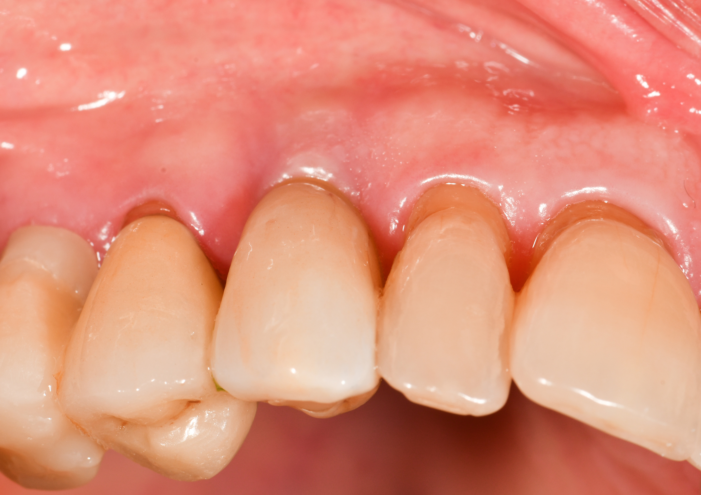
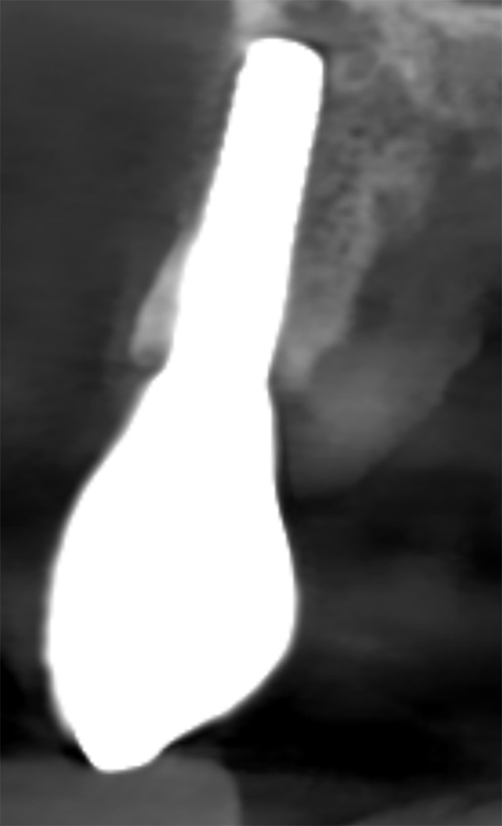
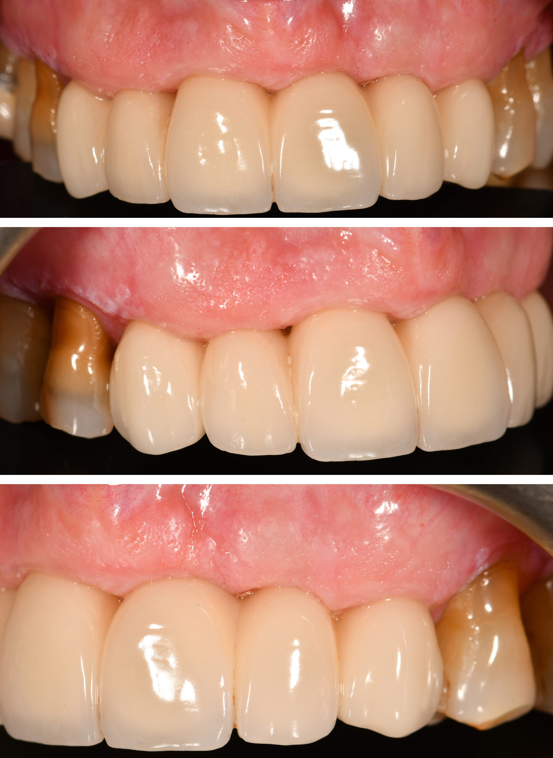
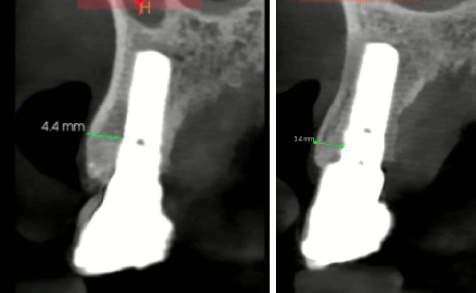
After 15 years in function, the clinical and radiographic images demonstrate excellent stability of the peri-implant tissues and marginal bone levels (Figs 9a – f). The CBCT scan taken after 15 years highlights that the palatally placed UR3 and UL3 have maintained a thick facial bone plate (3 – 4 mm) at the junction between the machined collar and the SLA surface (Figs 10a – c). It also shows bone coronal to the rough surface and, indeed, coronal to the implant-restoration margin. In contrast, the implant in #11 site is facially placed, and bone thickness is <1 mm (Fig. 10b). Had it been placed palatally, more facial bone would have been preserved (Fig. 10e). This case illustrates the correlation between facial bone and palatal positioning of the implant (Shahdad et al. 2023).

When not to use tissue level implants?

As shown, TL implants can be successfully used in the esthetic zone for IIP, achieving stable, long-term esthetic outcomes. We would argue that there are no contraindications for IIP specific to TL implants that do not also exist for BL implants. However, there are a few indications where TL implants are not favored over BL implants.

In the esthetic zone, a TL implant should be avoided: 

  • if the inter-tooth space is narrow so that <3 mm-diameter implant is required. This may be encountered with single lower incisors or upper lateral incisors and mesio-distally narrow pre-molars (bicuspids).
  • when replacing two adjacent anterior teeth and requiring two implants resulting in insufficient space (<4-mm inter-implant distance). In this case, should the occlusal scheme allow, a single TL implant with a cantilever pontic should be considered.

Immediate placement in sites with damaged facial socket bone carries a high risk of midfacial recession. Kan et al. (2007) demonstrated that 8.3% of immediate implants placed in sites with minor (V-shaped) facial dehiscence developed midfacial mucosal recession >1.5 mm one year later. This compared to 42.8% of sites with a moderately (U-shaped) large dehiscence (defect extended to the mesial or distal aspect) and 100% of sites with a large (UU-shaped) dehiscence (defect extended to the mesial or distal aspect of the adjacent teeth). Therefore, the extent of damage to the facial bone represents a significant risk factor for soft tissue recession, the risk increasing with the size of the dehiscence defect of the facial socket bone.

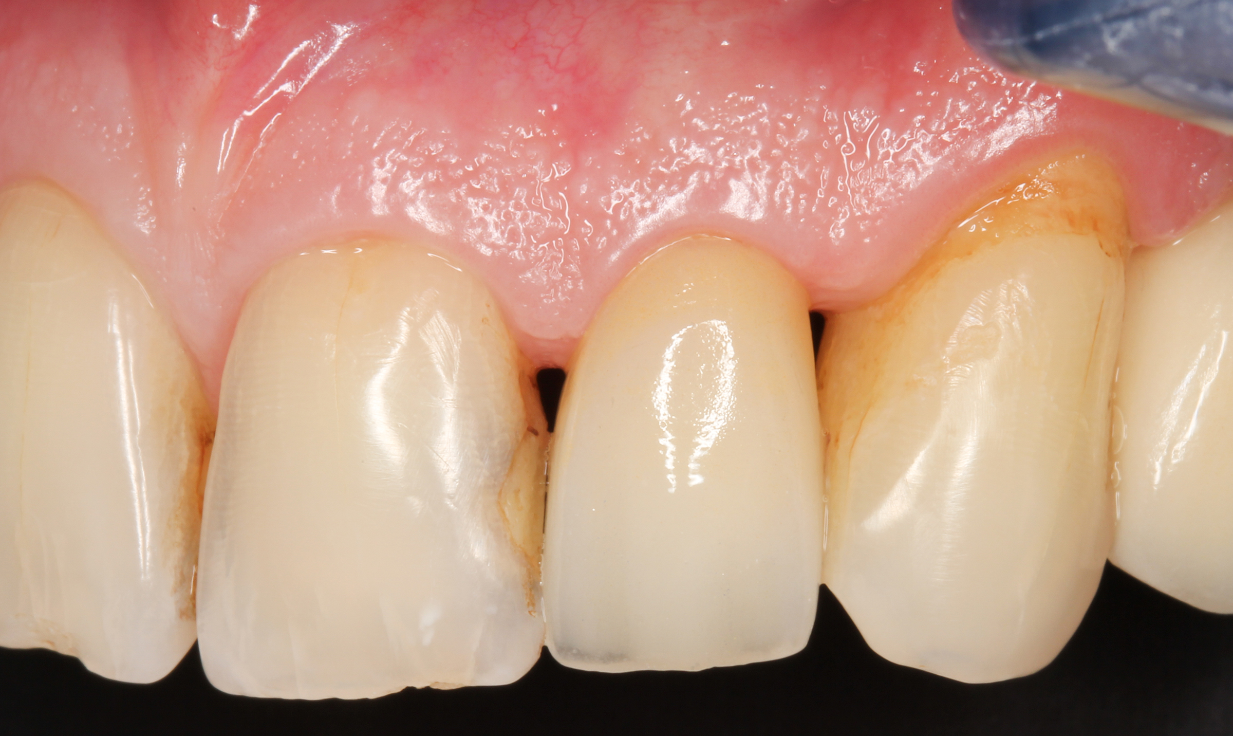
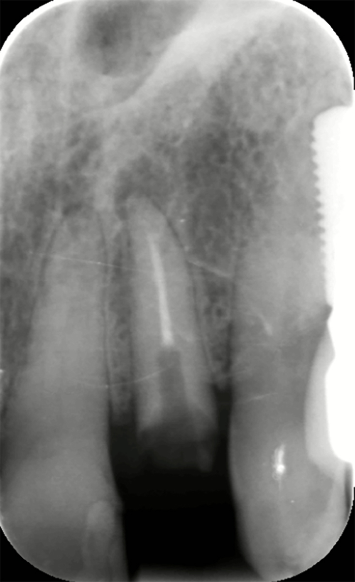
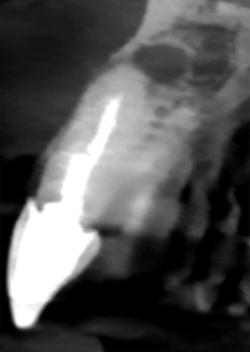
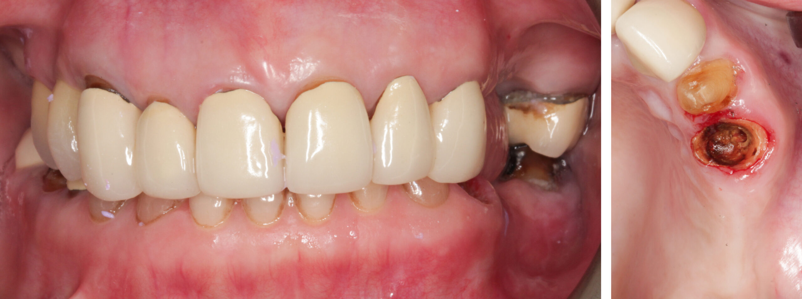
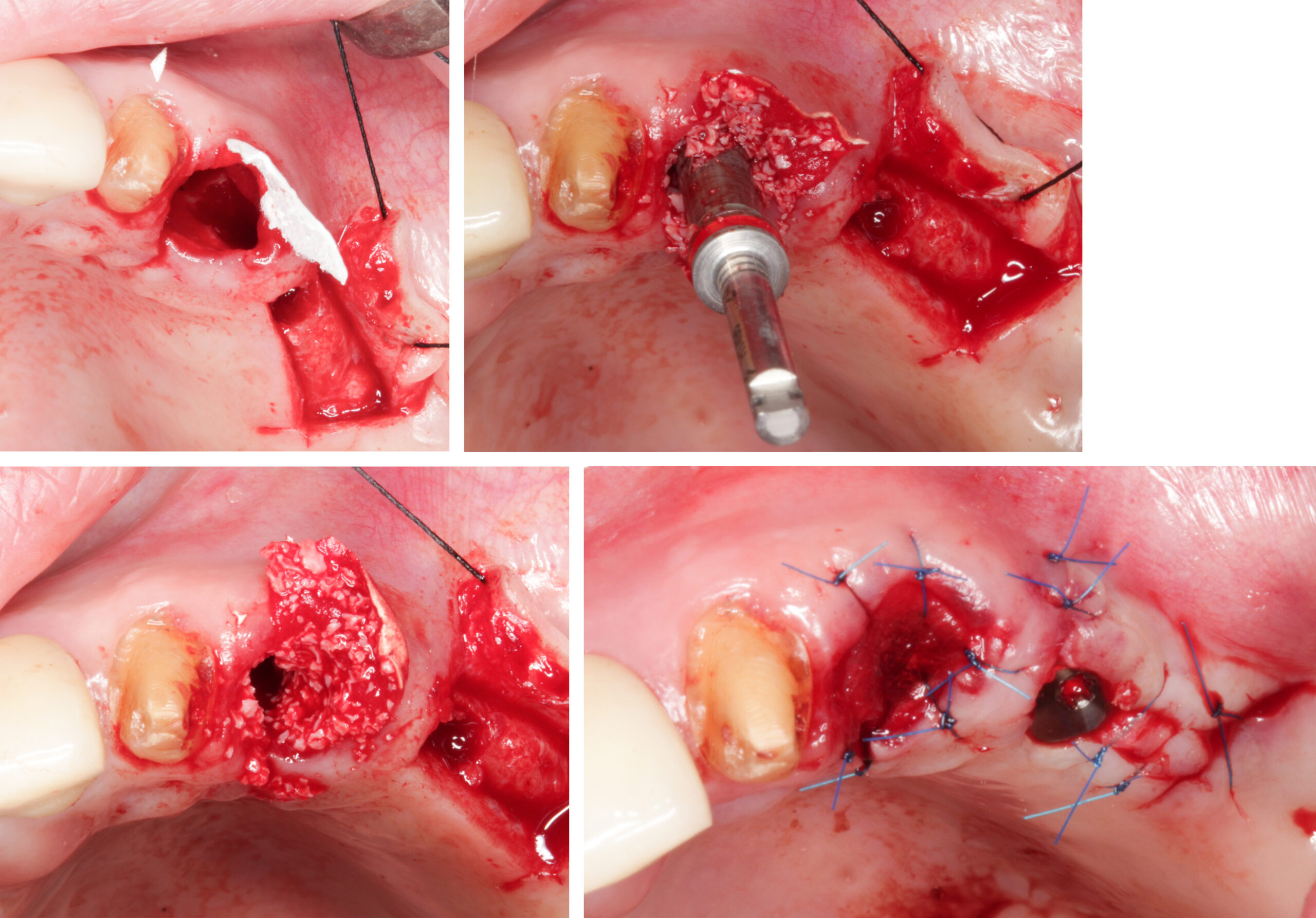
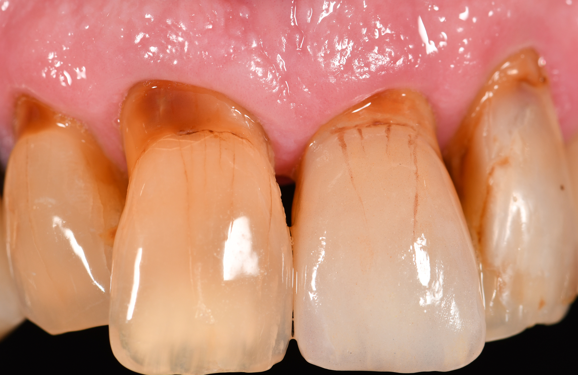
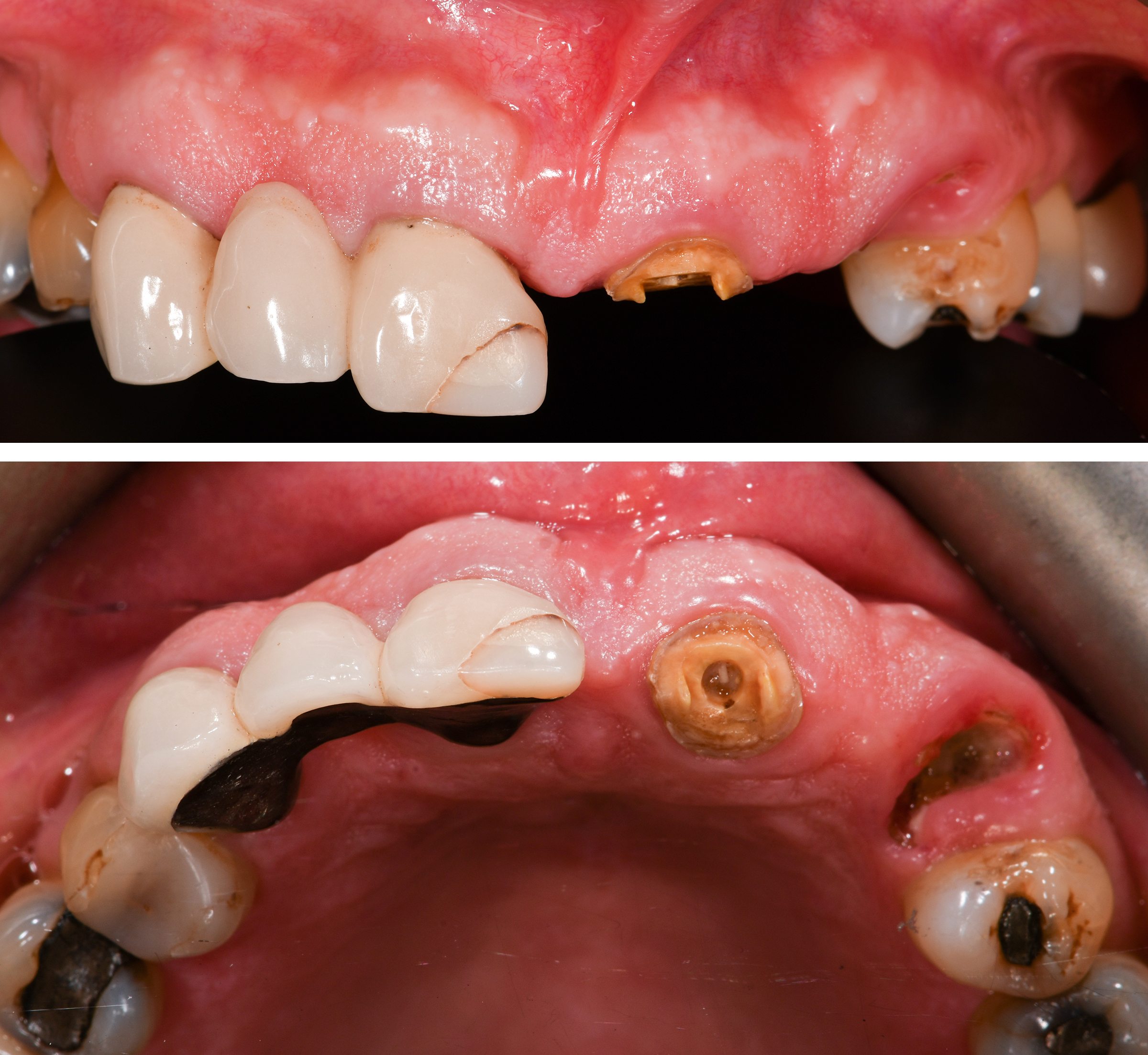
Let’s consider a very high-risk case of a 77-year-old female patient with a thin gingival phenotype (Fig. 11a). #23 post-core crown debonded, and the root was deemed unrestorable (Fig. 11b). The CBCT confirms Class IIB root position with <1 mm facial bone, if present (Fig. 11c). In pursuit of minimizing surgical morbidity in an elderly patient, an immediate implant in the #23 was decided upon in anticipation of encountering missing facial bone at placement.

Once the root was extracted, the facial plate was completely missing with a full root-length dehiscence. After osteotomy preparation, an internal socket flap was elevated along the three facial bone walls to allow for stabilization of a collagen membrane (Geistlich BioGide) between the flap and the facial bone (Fig. 11d). Particulate bone graft (Geistlich BioOss) was positioned and shaped with the implant drill to replace the missing facial bone (Figs 11e – f).

A Straumann Tissue Level Tapered Effect (TE) – a coronally tapered implant with “active thread” designed for higher primary stability – was placed in the socket. At the same time, a standard TL implant was placed in the healed ridge to replace the bicuspids (#24, #25) (Fig. 11g). The membrane was wrapped on top of the 2-mm bevelled healing abutment.

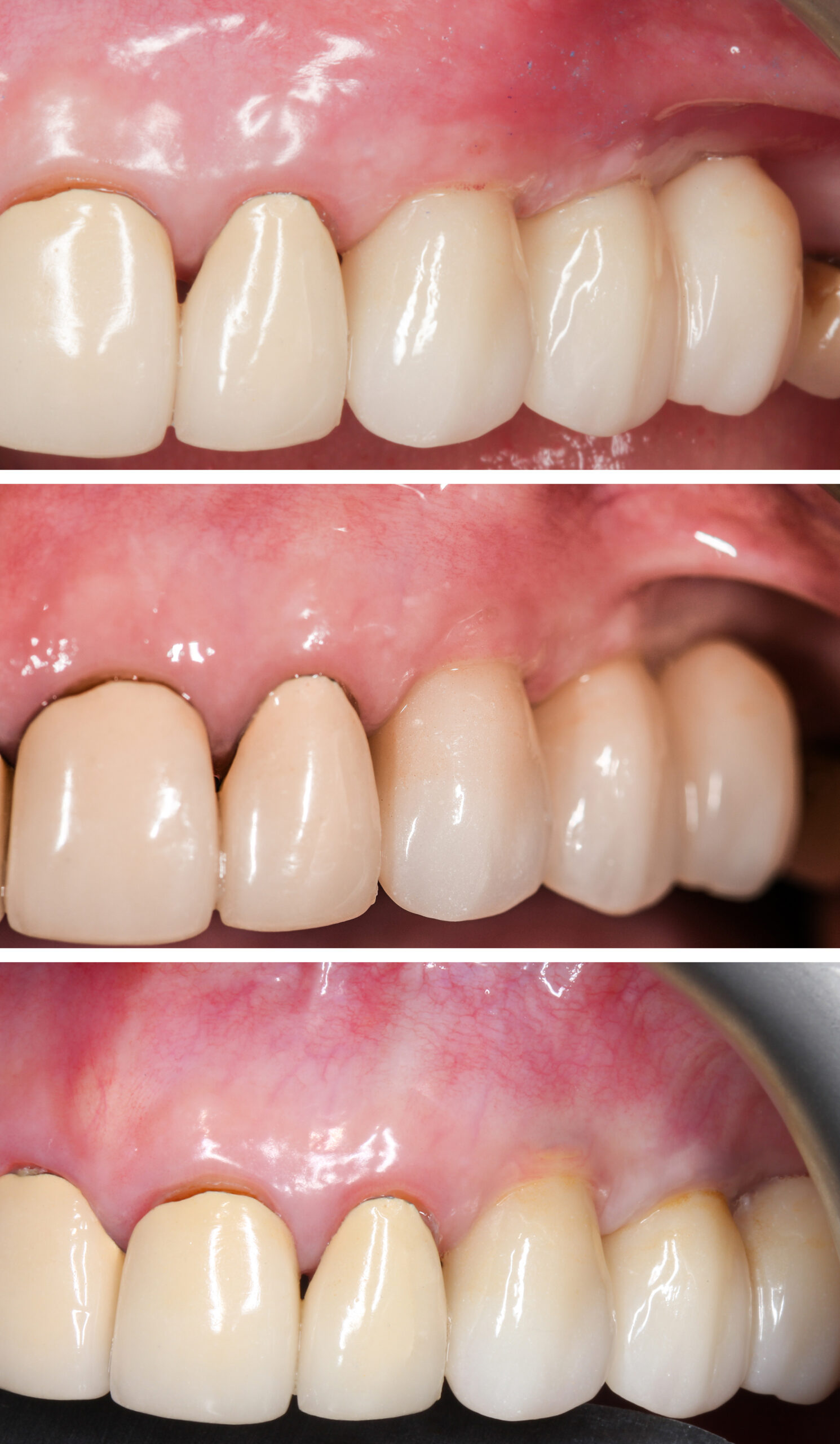
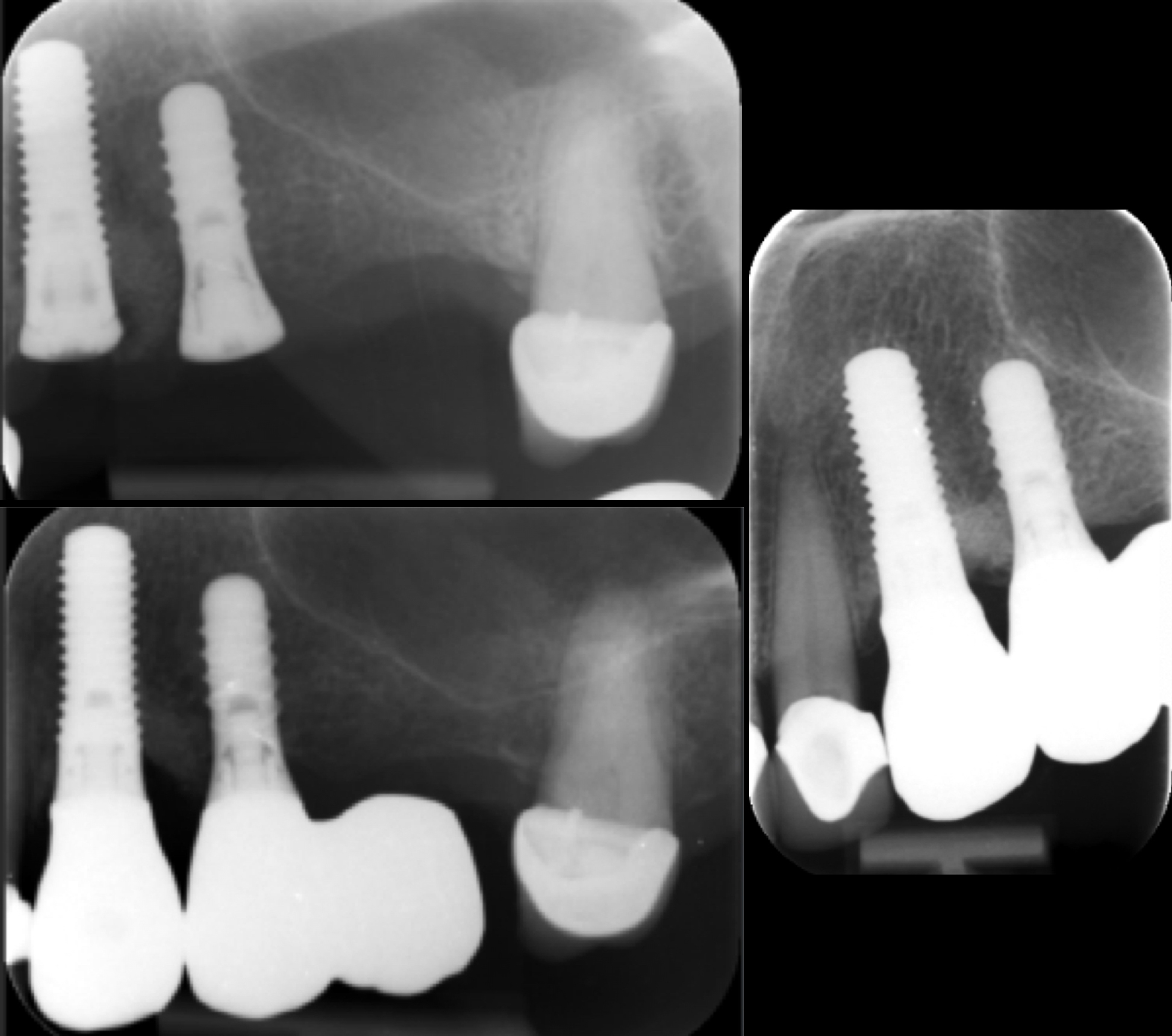
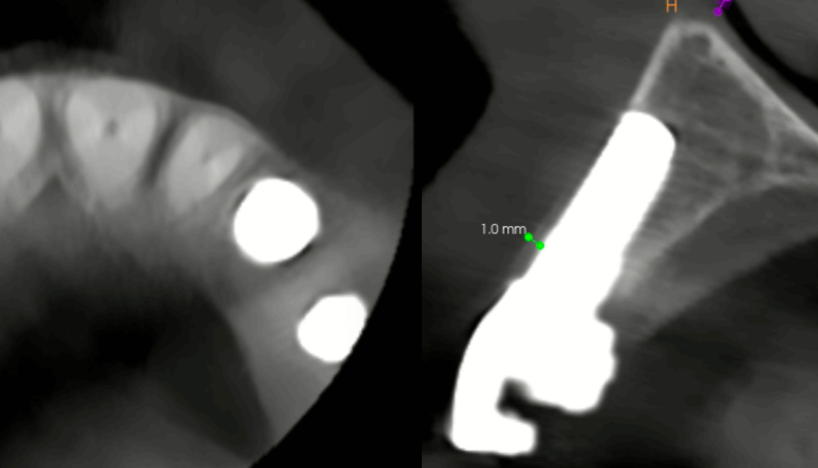
Immediate post-restoration, 1-year and 6-year reviews (Fig. 11h – j) demonstrate that the mucosal zenith had stabilized by the end of 1 year and remained stable over the next 5 years, which is also confirmed radiographically (Figs 11k – m). Despite a complete lack of pre-surgical facial bone in a thin gingival phenotype, an excellent PES score was achieved in contrast to those reported by Kan et al. (2007). The CBCT scan after 6 years shows that the buccal bone reconstruction yielded limited success with <1 mm bone thickness (Figs 11n – o). Again, a more palatally positioned implant could have achieved a better outcome; a challenge clinicians face when trying to control the implant position, especially with a coronally tapered implant design.

Whilst this case demonstrates a successful result in a very high-risk case, we would caution against such an aggressive Type 1 protocol, as a large U-shaped or UU shaped bone defect is contraindicated for immediate placement. An early implant placement protocol would have achieved a more predictable bone regeneration.

Soft tissue considerations (gingival phenotype and deficiencies)

A thin phenotype increases the risk of recession and compromised soft tissue response following surgical or restorative therapy (Romandini et al. 2021). Immediate implants placed in sites with a thin gingival phenotype are associated with significantly more recession of the midfacial mucosa than sites with a thick gingival phenotype (Kan et al. 2003; Kan et al., 2011).

A recent systematic review concluded that the placement of a connective tissue graft contributes to midfacial soft tissue stability following immediate placement and should be considered when an elevated risk for midfacial recession (thin soft tissue phenotype, < 0.5-mm facial bone thickness) is expected in the esthetic zone (Seyssens et al. 2021).

The risks can be similar for TL implants, but the authors have not experienced increased mucosal recession after placing TL implants in similar high-risk patients. Fig. 12 shows a 69-year-old male patient with subgingival root caries in maxillary left central incisor (#21).

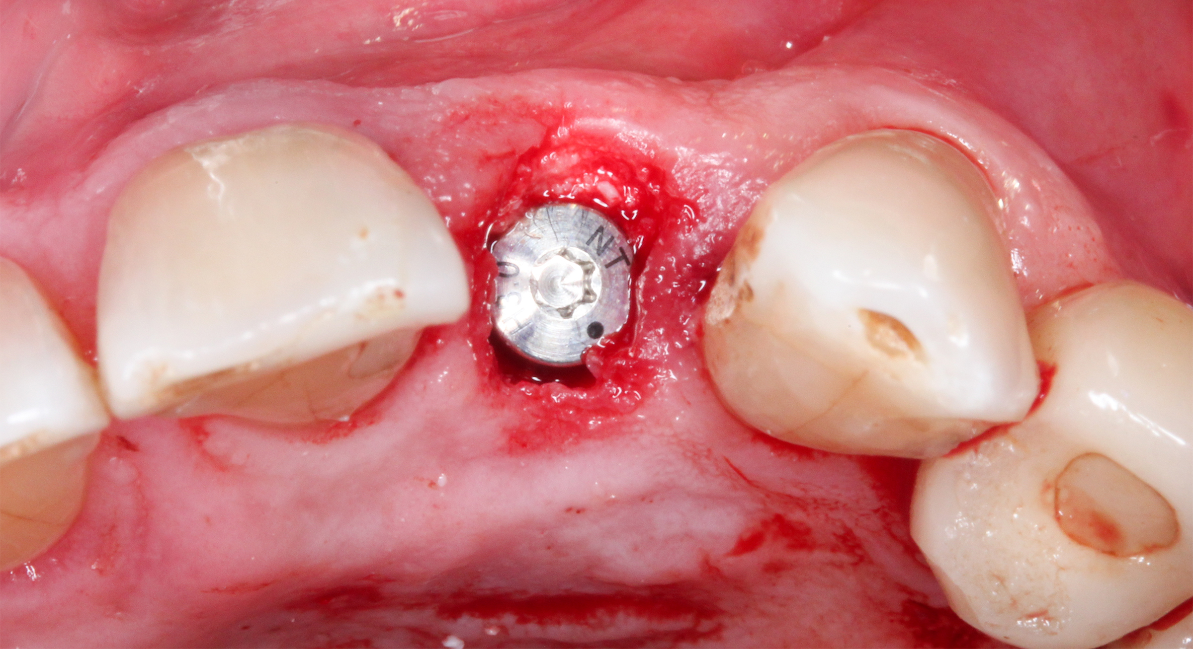
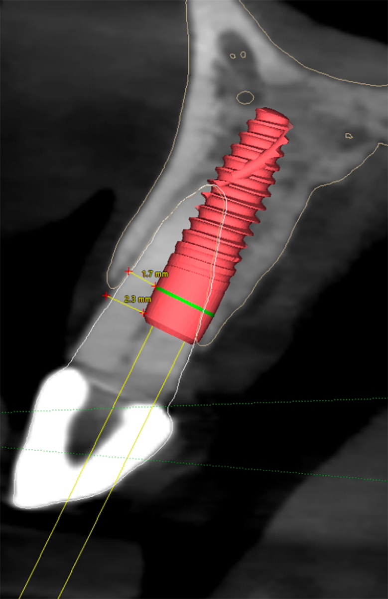
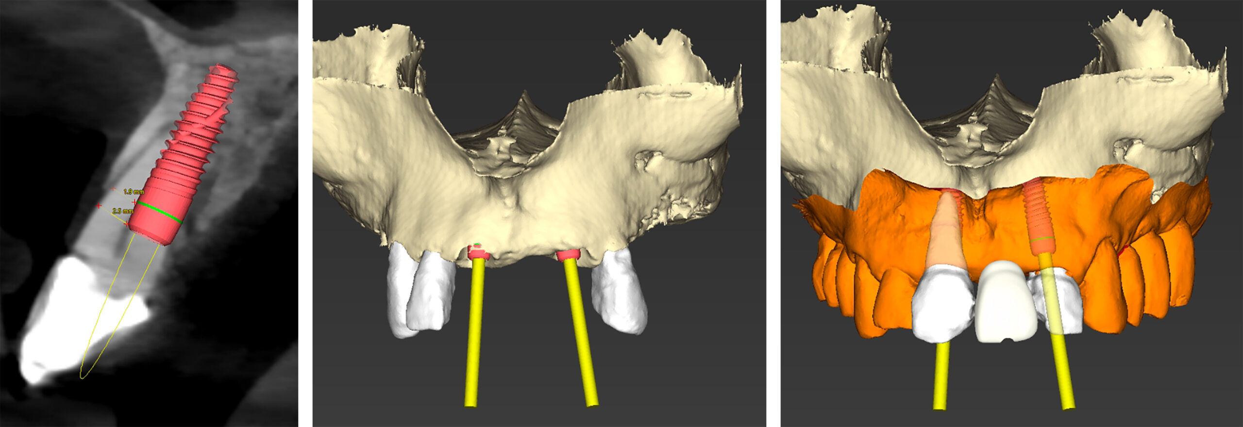
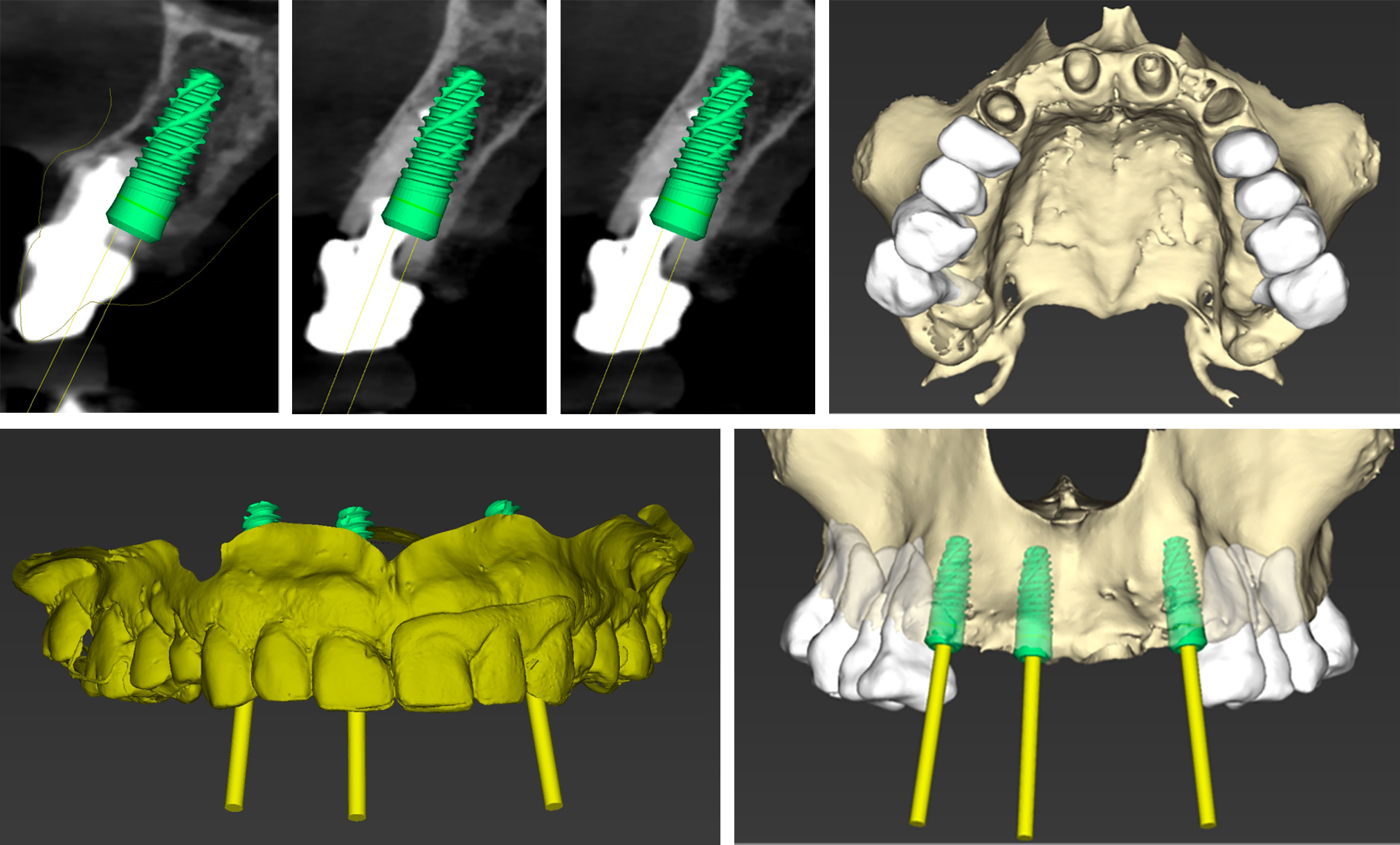
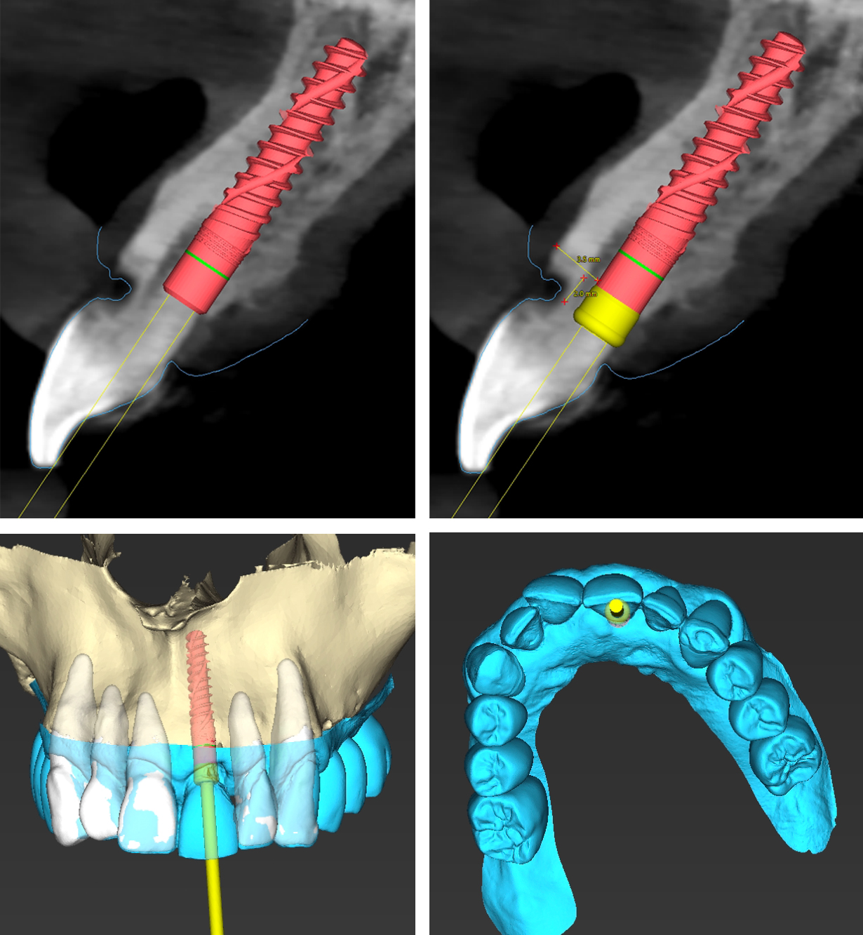
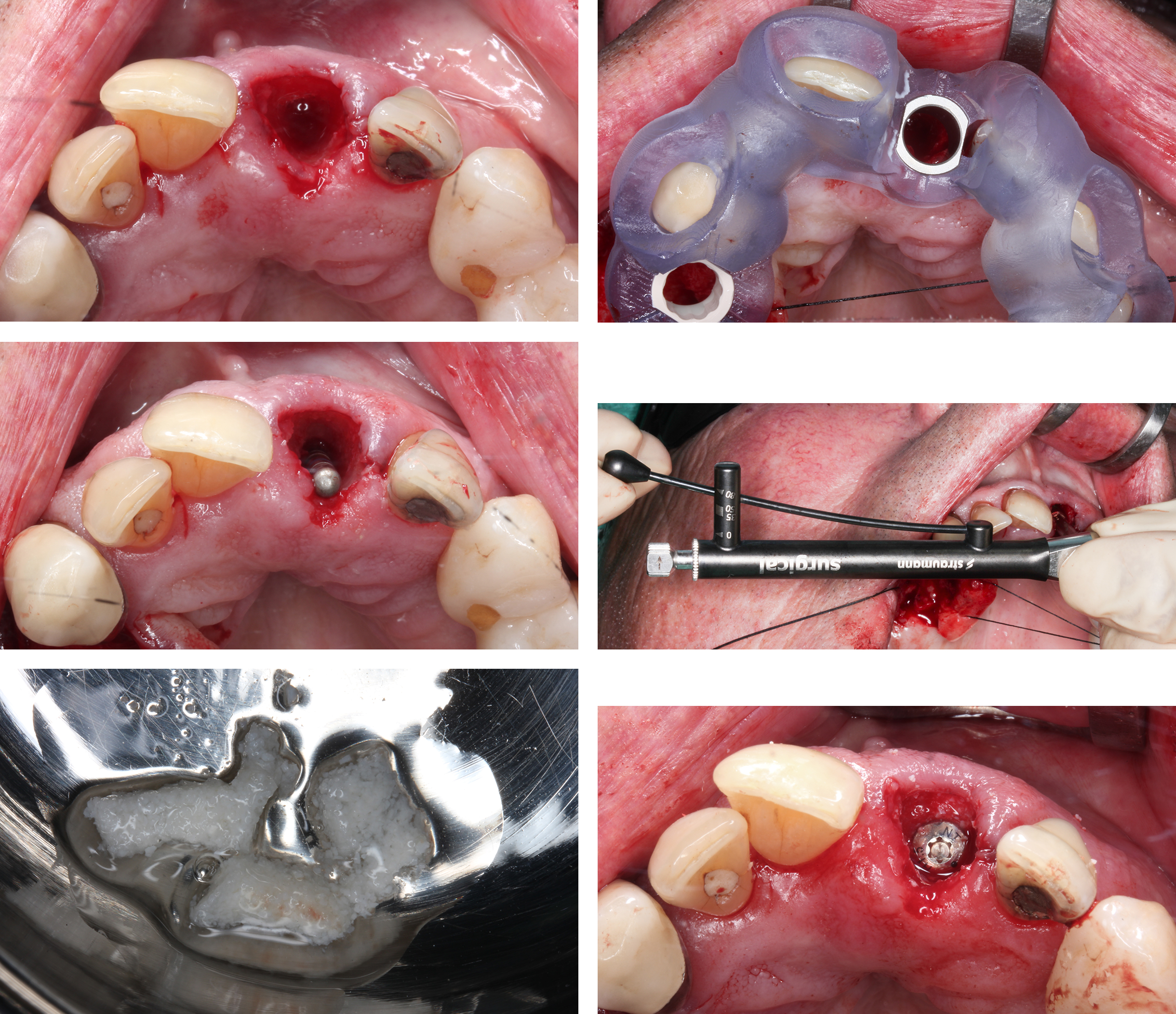
There is a pre-existing gingival recession, and the gingival margin of #21 is apical to #11 (Fig. 12a). The CBCT scan shows class III root position and <1 mm buccal bone thickness with a vertical bone loss up to 50% of the root length (Figs 12b – c). There was no evidence of peri-apical pathology. A fully-guided implant protocol was used for flapless placement (Figs 12 d – i) and immediate provisionalization. The implant axis was planned palatal to the root axis (Fig. 12j).

A fully tapered TL implant (Straumann TLX) for high primary stability (Fig. 12k) with NT collar design was selected to allow a narrower emergence angle to mimic recession in the implant crown. The horizontal bone defect was filled with DBBM-C soaked in Regenfast (hyaluronic acid) (Figs 12l – m). A titanium temporary abutment was attached (Fig. 12n) and a 3D-printed PMMA provisional crown was used for chairside pick-up with dual cure carbon-fiber reinforced composite resin (Build-It™, Pentron, USA). An undercontoured, concave labial soft tissue support was provided for immediate provisional restoration (Fig. 12o – p).

After 1 week, follow-up showed excellent soft tissue response and evidence of coronal growth rather than apical migration of the mucosal margin (Fig. 12q). Modifications of the provisional were carried out to optimize the emergence contours (Fig. 12r). The definitive restoration on a Ti-base abutment using a zirconia coping with facial ceramic veneering was provided as the definitive crown (Figs 12s – t).

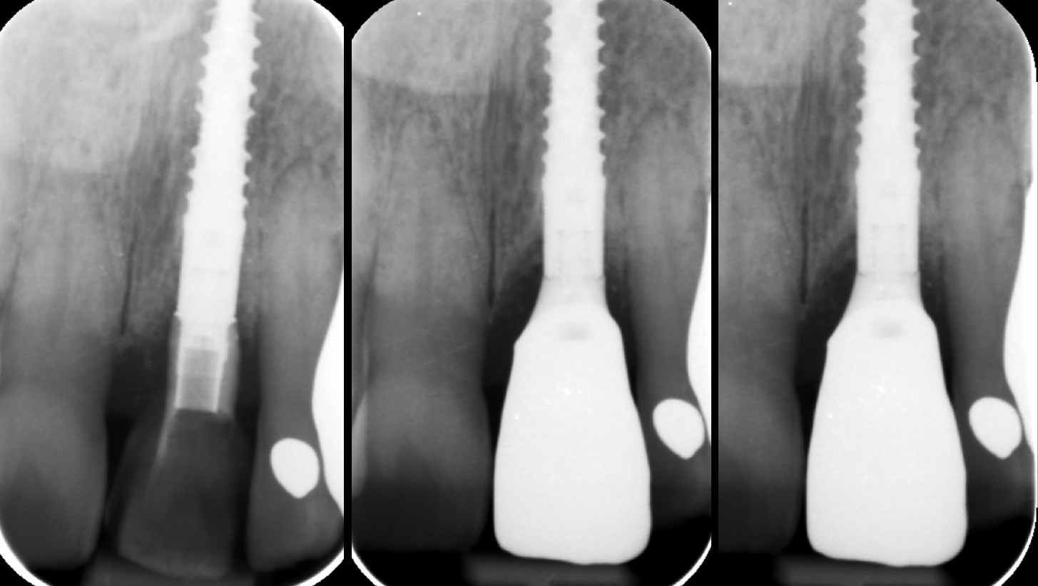
An 18-month follow-up showed stable peri-implant soft tissues and an excellent esthetic result. The peri-apical radiograph also confirmed stable marginal bone levels (Fig. 12u) and clinically, the mucosal zenith of #21 was at least 1 mm coronal to #11 (Fig. 12v) in contrast to the preoperative situation when recession in #21 was apical to #11.

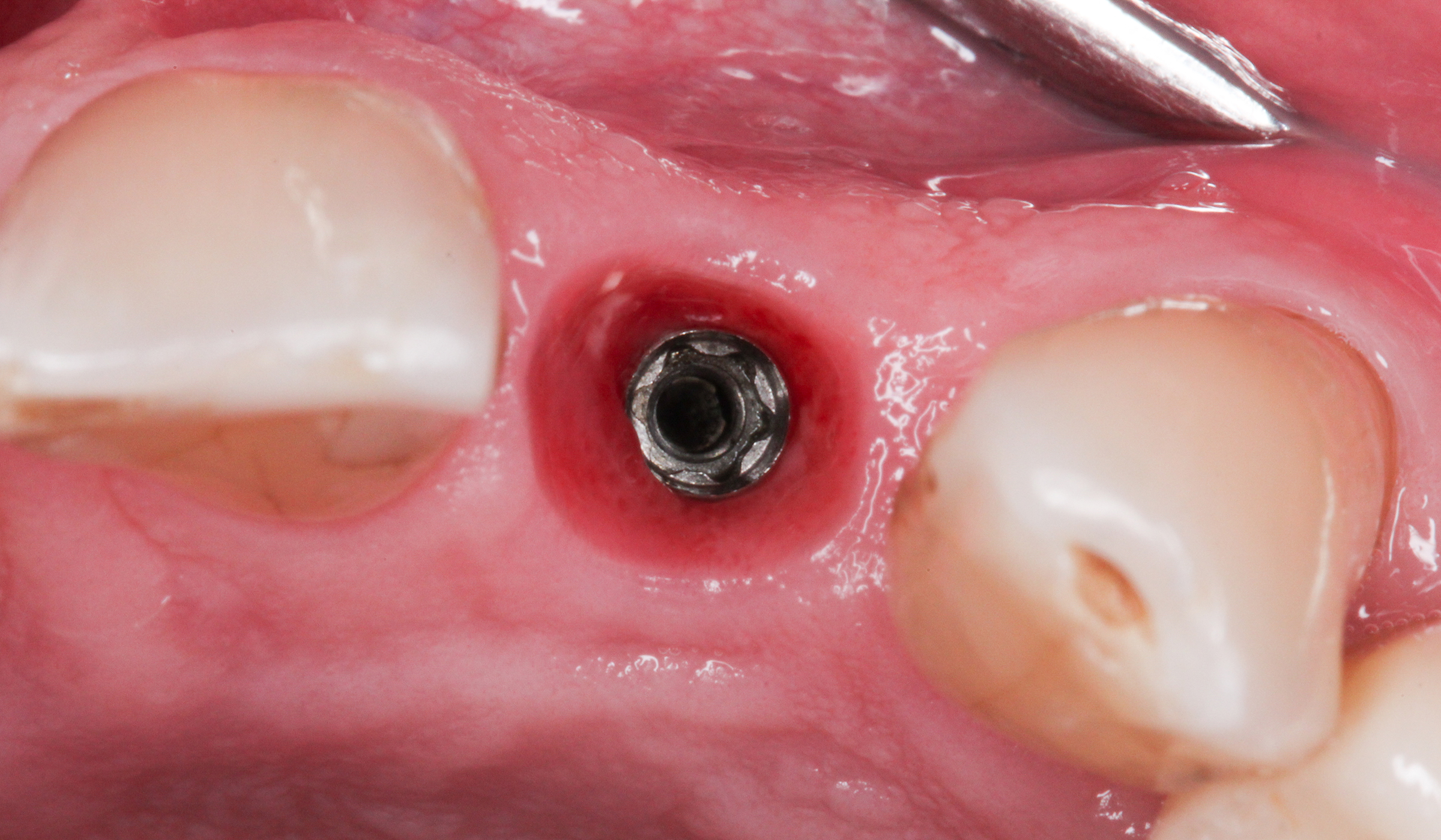
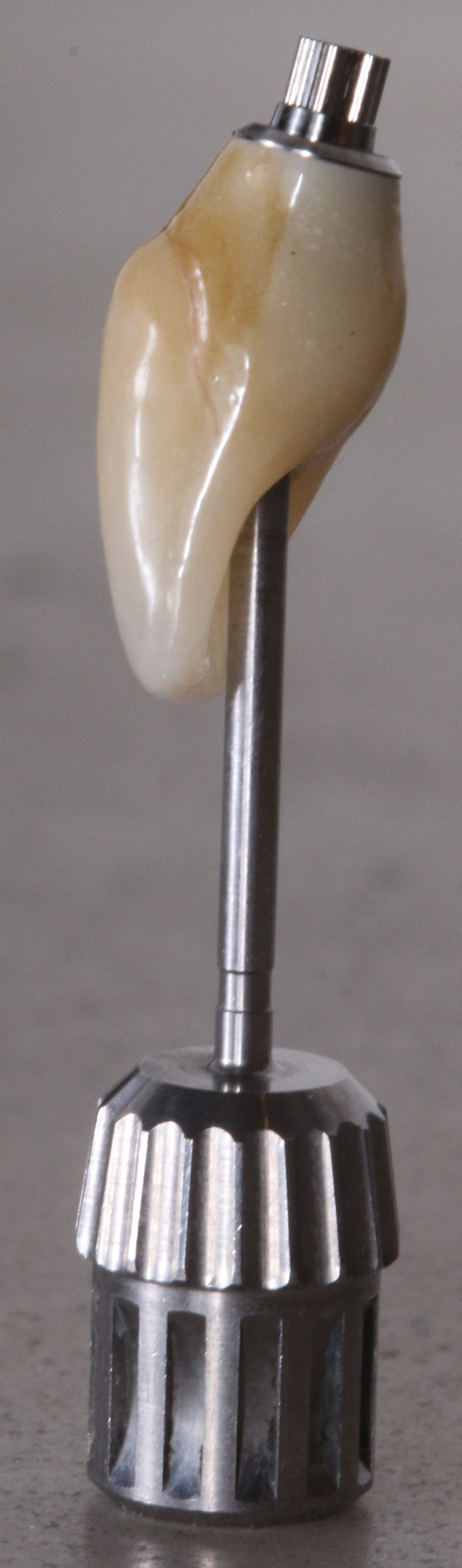
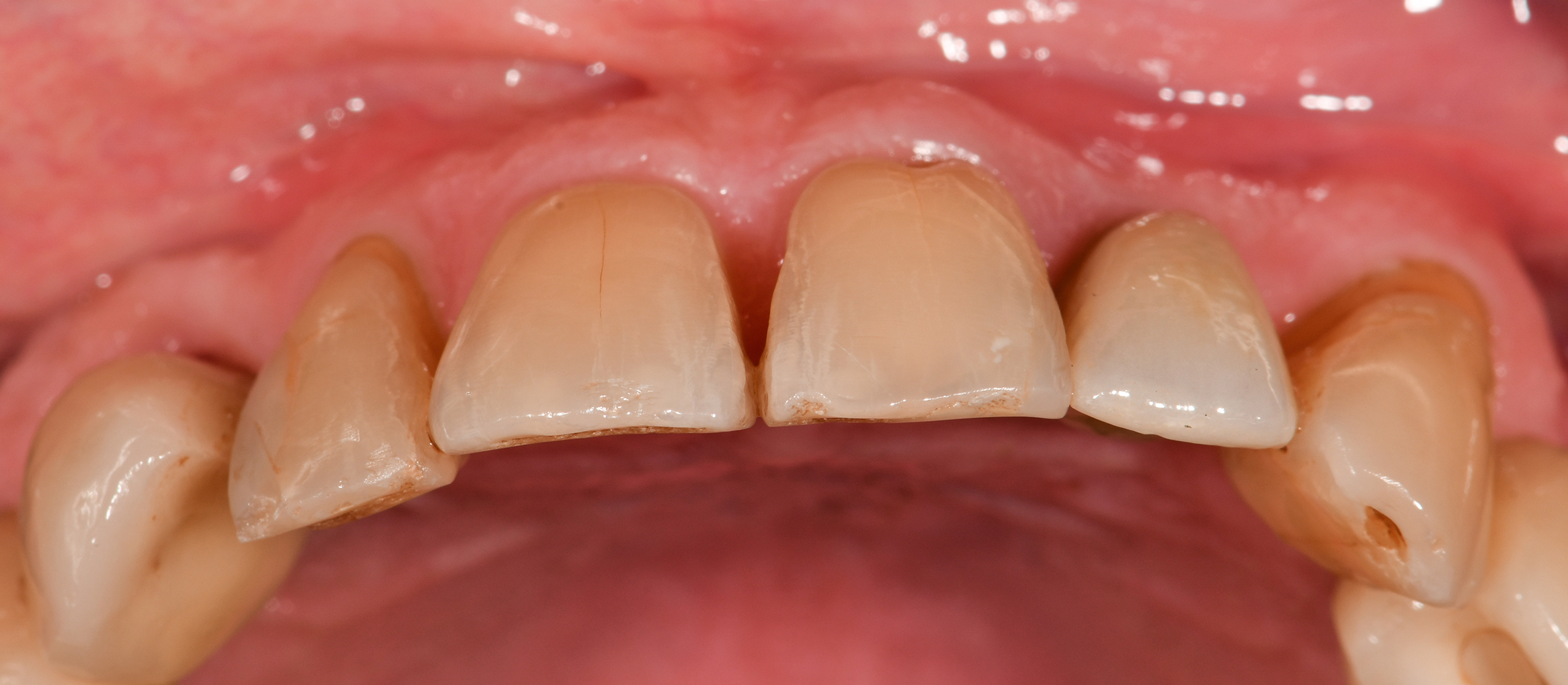
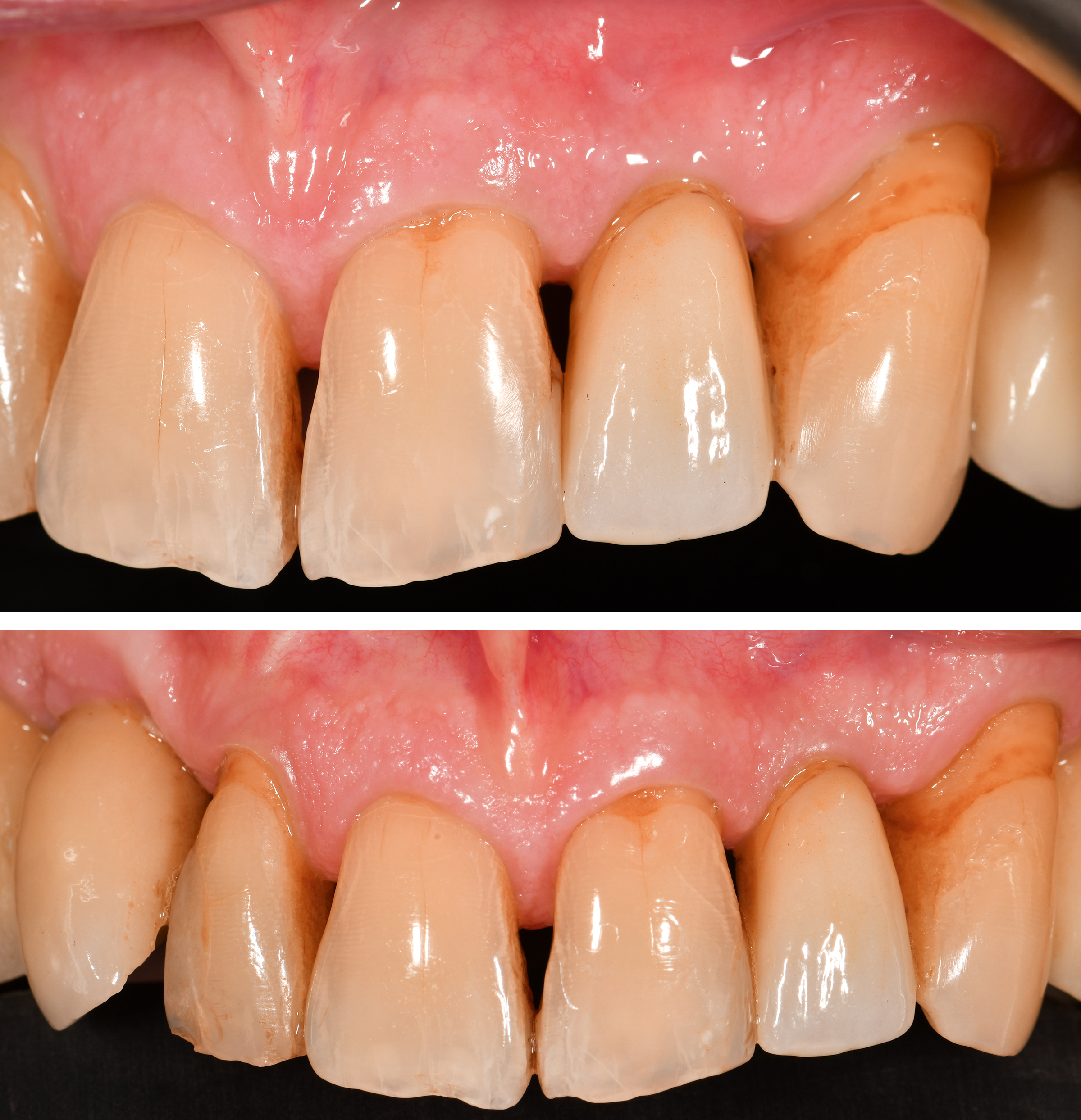
The recent introduction of an apically tapered TL implant (Straumann TLC) allows for a much more controlled implant positioning in extraction sockets whilst achieving high primary stability and complementing the benefits of the machined collar for simultaneous osseo- and mucointegration.

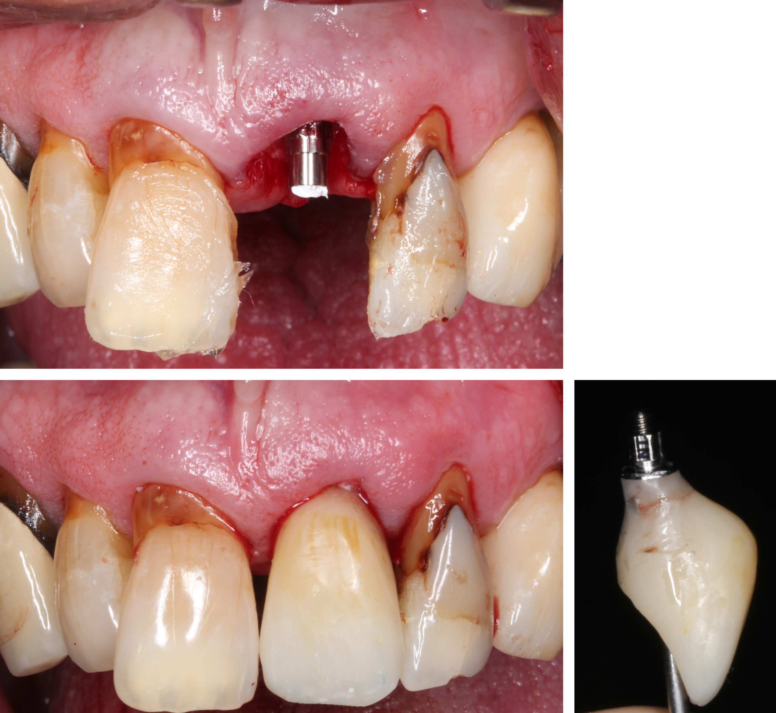
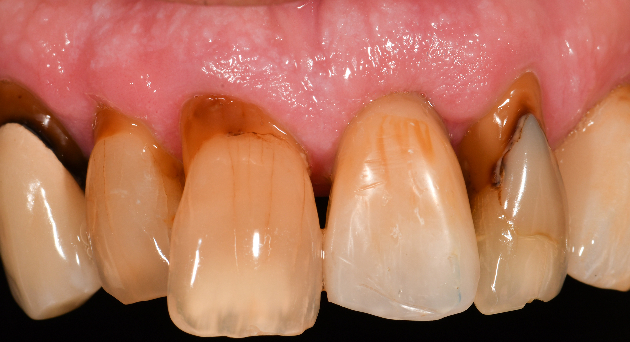
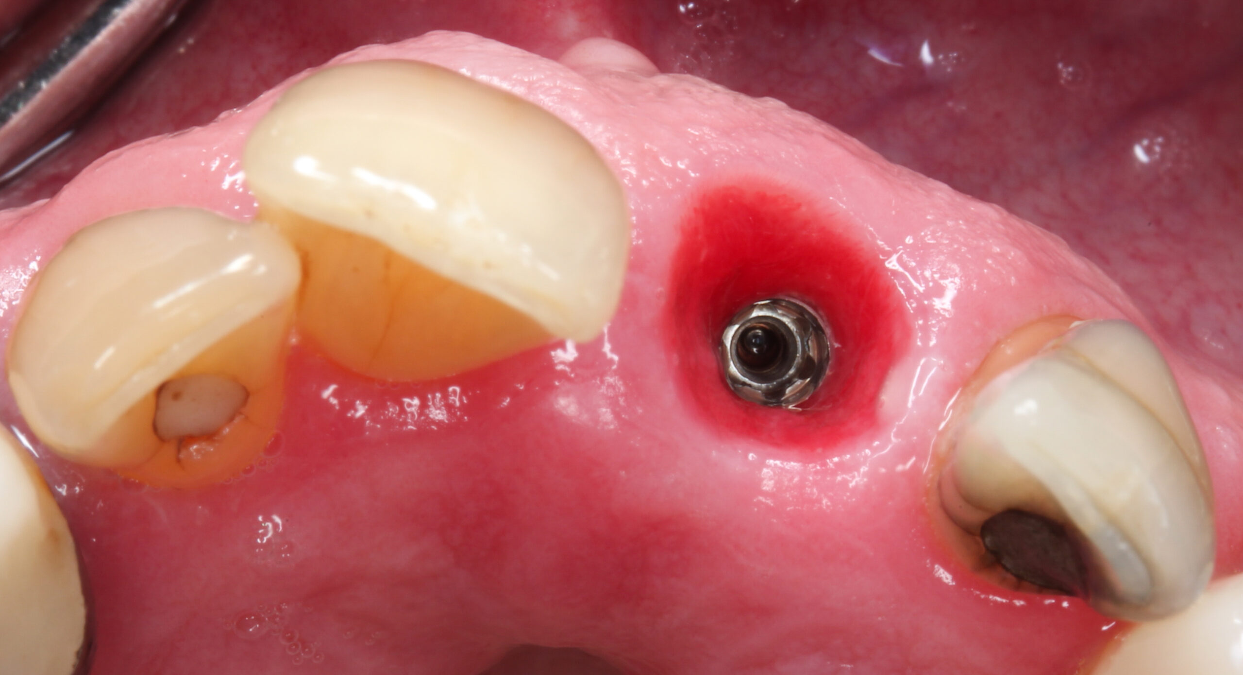
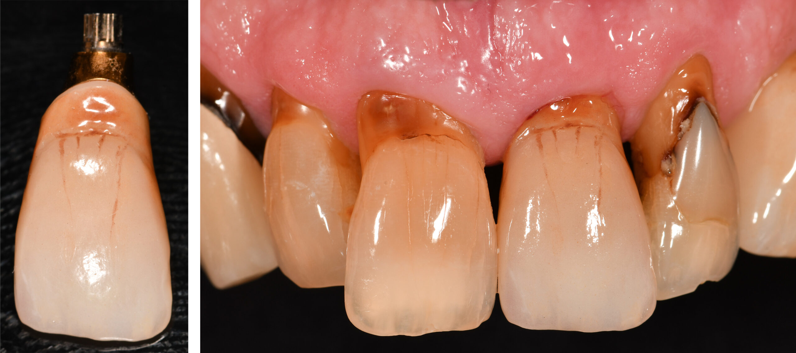
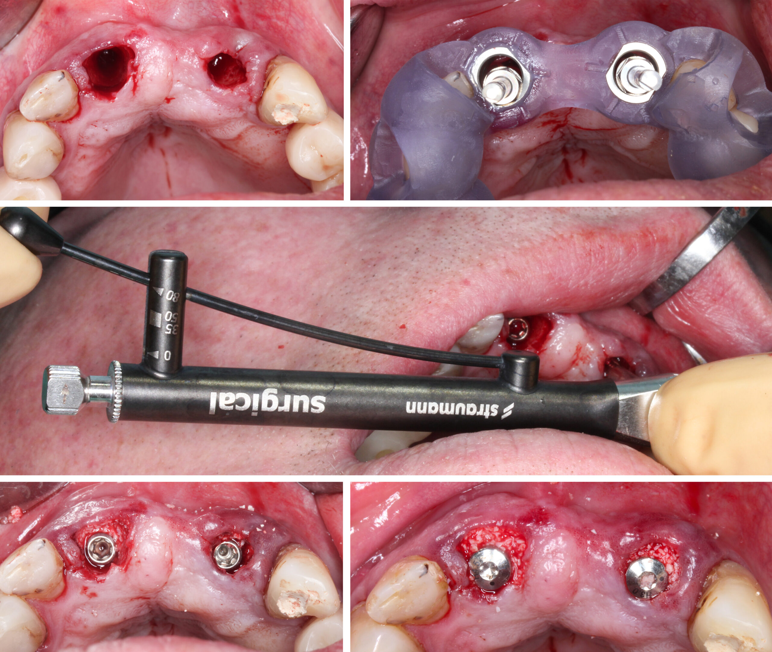
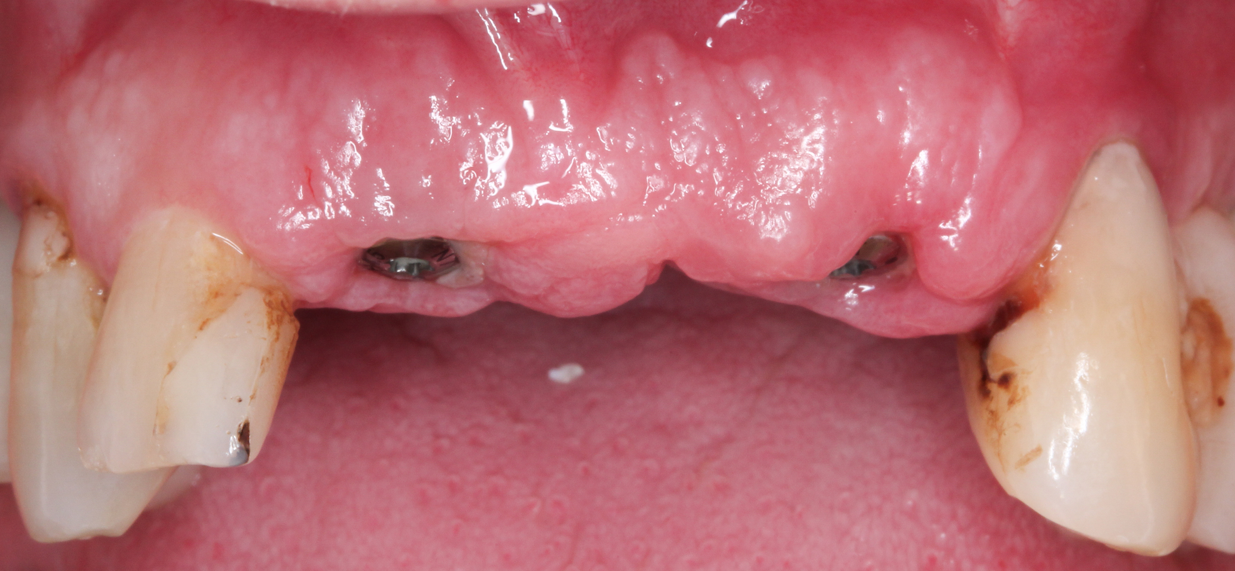
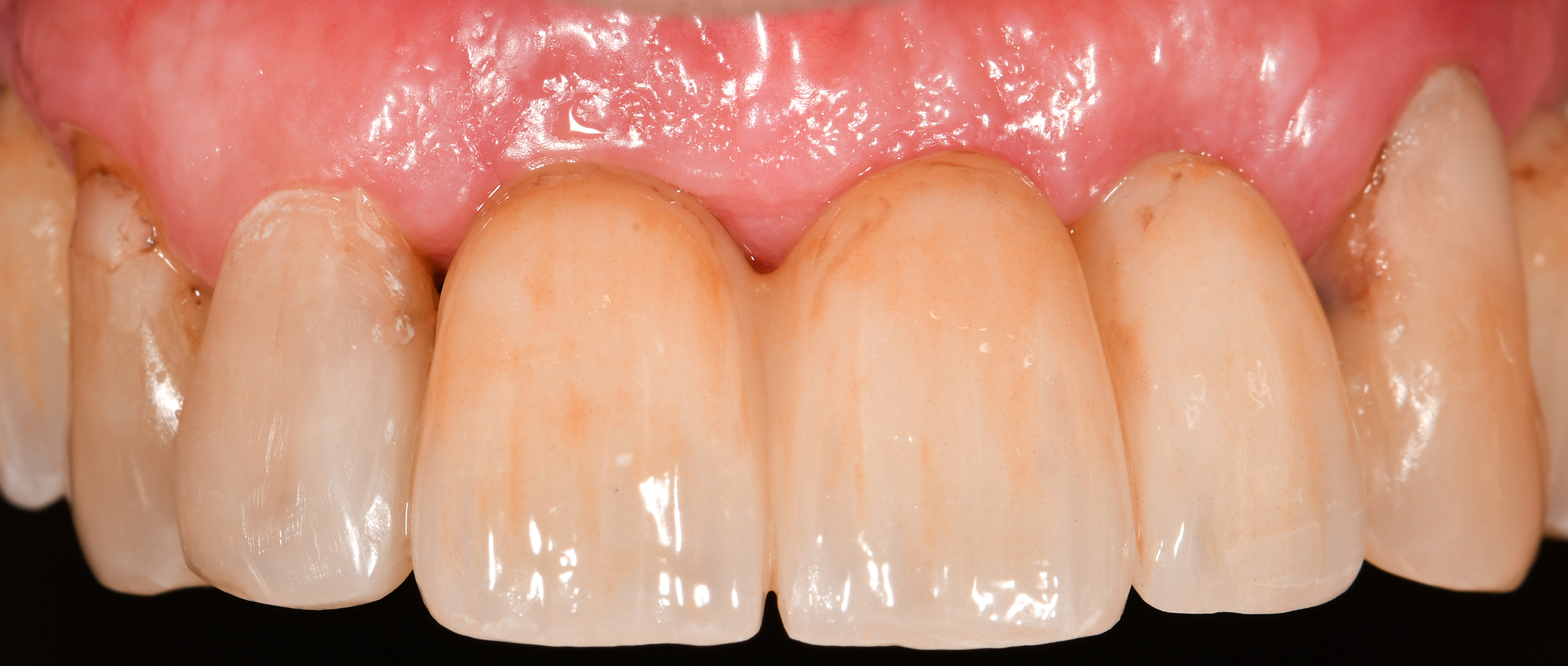
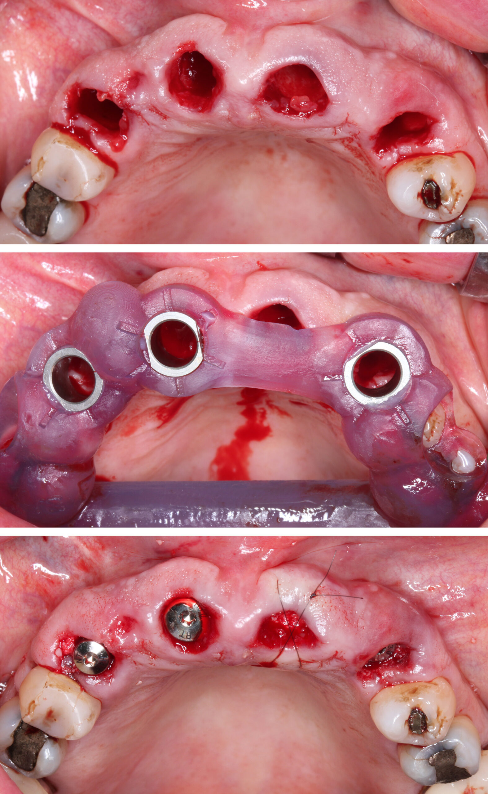
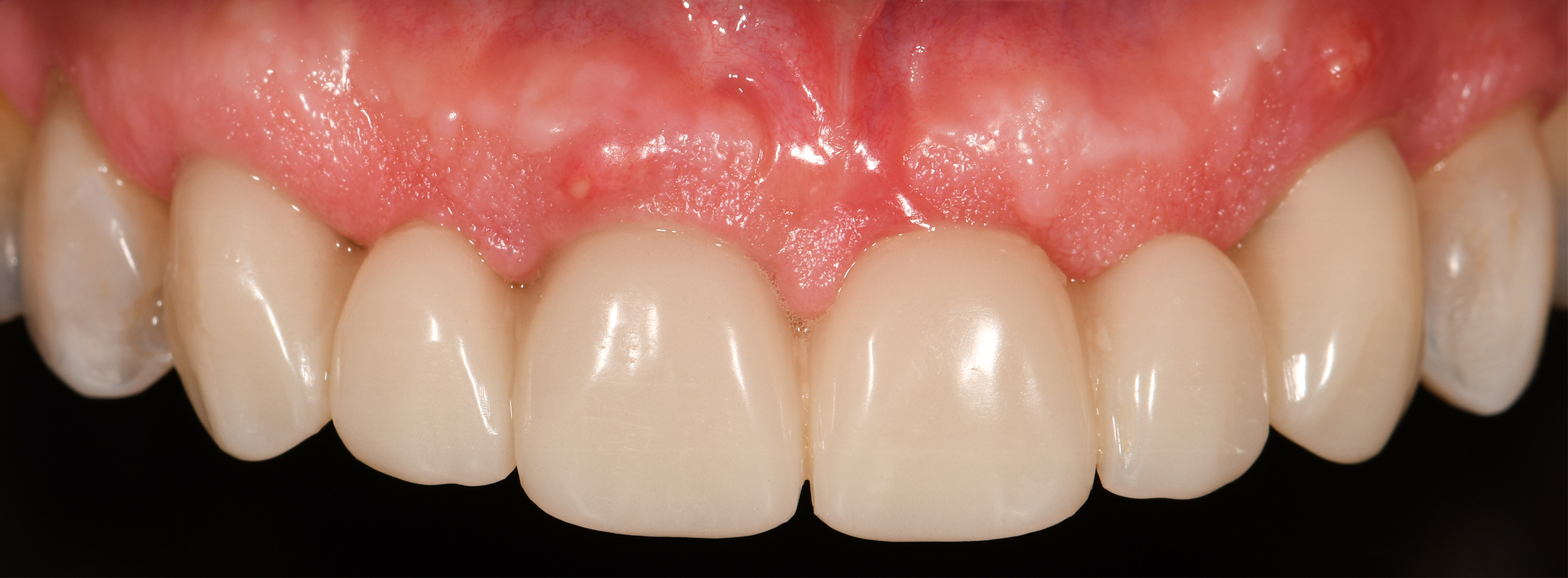
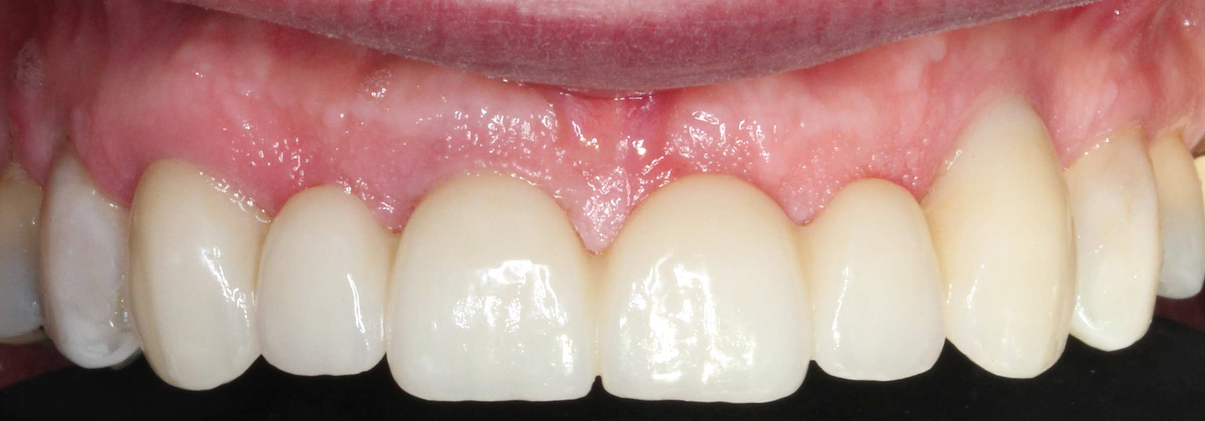
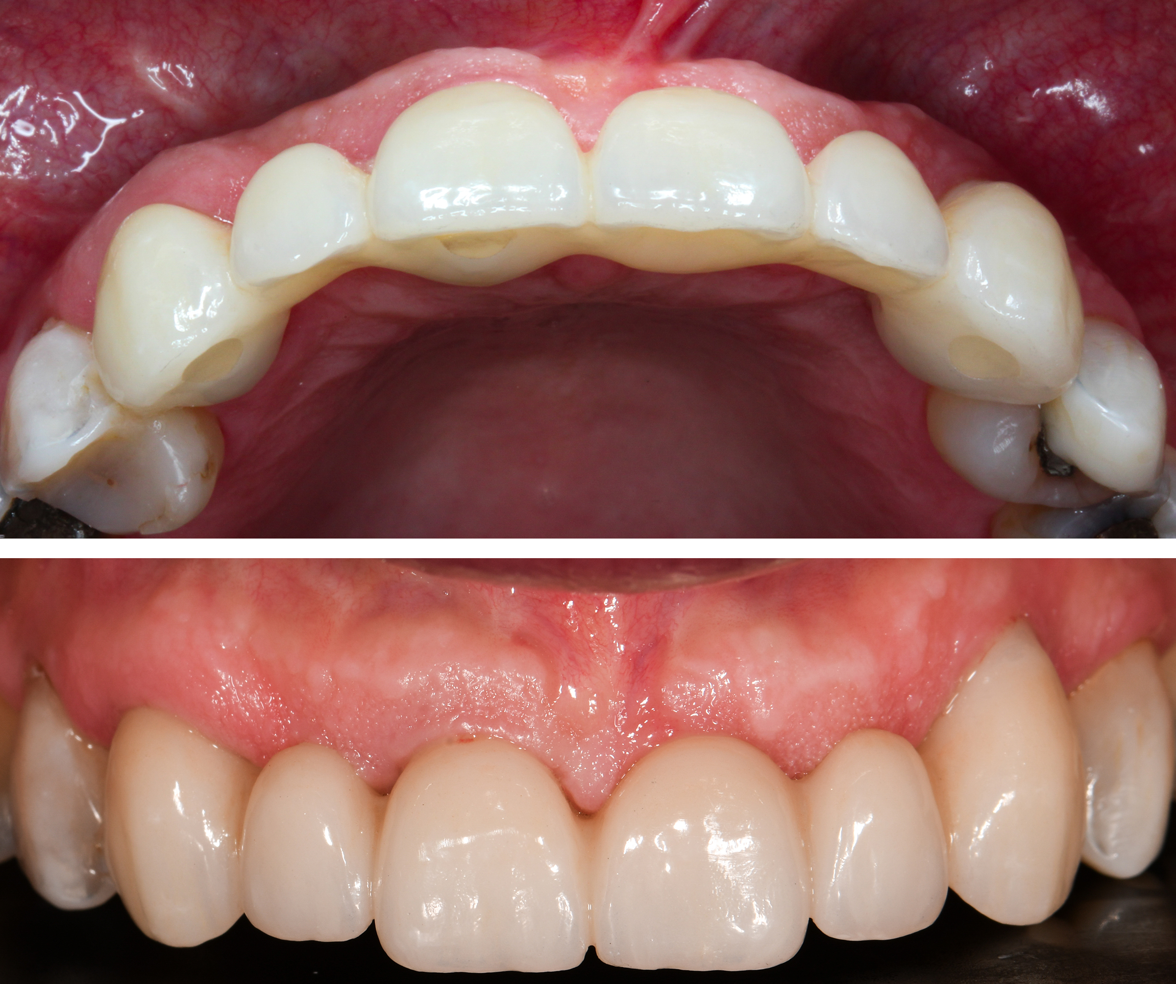
We conclude this article demonstrating two maxillary anterior cases treated with apically tapered TL implants (Figs 13 and 14).

Concluding Remarks

Proper case selection for IIP with TL implants is essential and correct 3D positioning of the implant is required to achieve stable esthetic outcomes. Deviation from this position is likely to result in suboptimal esthetic outcomes. No soft tissue grafting was carried out in any of the cases. Nevertheless, the pink esthetics achieved were optimal with healthy, pink mucosa. There was no shine-through of the TL collar, which is often proposed as a complication and a reason for not choosing TL implants in an esthetic zone. TL implants with tapered geometries (e.g, Straumann TLX and Straumann TLC) aid in achieving predictable primary stability for IIP. Therefore, the TL implant can be utilized predictably in the esthetic zone, and modern apically tapered geometries provide excellent primary stability for IIP.

Abrahamsson I, Berglundh T, Lindhe J. The mucosal barrier following abutment dis/reconnection. An experimental study in dogs. J Clin Periodontol. 1997 Aug;24(8):568-72. doi: 10.1111/j.1600-051x.1997.tb00230.x. PMID: 9266344.

Akkocaoglu M, Uysal S, Tekdemir I, Akca K, Cehreli MC. Implant design and intraosseous stability of immediately placed implants: a human cadaver study. Clin Oral Implants Res. 2005 Apr;16(2):202-9. doi: 10.1111/j.1600-0501.2004.01099.x. PMID: 15777330.

Alves CC, Muñoz F, Cantalapiedra A, Ramos I, Neves M, Blanco J. Marginal bone and soft tissue behavior following platform switching abutment connection/disconnection–a dog model study. Clin Oral Implants Res. 2015 Sep;26(9):983-91. doi: 10.1111/clr.12385. Epub 2014 Apr 16. PMID: 24735458.

Andersen E, Haanaes HR, Knutsen BM. Immediate loading of single-tooth ITI implants in the anterior maxilla: a prospective 5-year pilot study. Clin Oral Implants Res. 2002 Jun;13(3):281-7. doi: 10.1034/j.1600-0501.2002.130307.x. PMID: 12010158.

Araújo MG, Lindhe J. Dimensional ridge alterations following tooth extraction. An experimental study in the dog. J Clin Periodontol. 2005 Feb;32(2):212-8. doi: 10.1111/j.1600-051X.2005.00642.x. PMID: 15691354.

Atieh MA, Ibrahim HM, Atieh AH. Platform switching for marginal bone preservation around dental implants: a systematic review and meta-analysis. J Periodontol. 2010 Oct;81(10):1350-66. doi: 10.1902/jop.2010.100232. PMID: 20575657.

Benic GI, Mokti M, Chen CJ, Weber HP, Hämmerle CH, Gallucci GO. Dimensions of buccal bone and mucosa at immediately placed implants after 7 years: a clinical and cone beam computed tomography study. Clin Oral Implants Res. 2012 May;23(5):560-6. doi: 10.1111/j.1600-0501.2011.02253.x. Epub 2011 Sep 8. PMID: 22093013.

Berglundh T, Lindhe J. Dimension of the periimplant mucosa. Biological width revisited. J Clin Periodontol. 1996 Oct;23(10):971-3. doi: 10.1111/j.1600-051x.1996.tb00520.x. PMID: 8915028.

Bianchi AE, Sanfilippo F. Single-tooth replacement by immediate implant and connective tissue graft: a 1-9-year clinical evaluation. Clin Oral Implants Res. 2004 Jun;15(3):269-77. doi: 10.1111/j.1600-0501.2004.01020.x. PMID: 15142088.

Brägger U, Hämmerle CH, Lang NP. Immediate transmucosal implants using the principle of guided tissue regeneration (II). A cross-sectional study comparing the clinical outcome 1 year after immediate to standard implant placement. Clin Oral Implants Res. 1996 Sep;7(3):268-76. doi: 10.1034/j.1600-0501.1996.070309.x. PMID: 9151591.

Brånemark PI, Adell R, Breine U, Hansson BO, Lindström J, Ohlsson A. Intra-osseous anchorage of dental prostheses. I. Experimental studies. Scand J Plast Reconstr Surg. 1969;3(2):81-100. doi: 10.3109/02844316909036699. PMID: 4924041.

Bressan E, Guazzo R, Tomasi C, Peña TG, Galindo-Moreno P, Caponio VCA, Sbricoli L, Canullo L. Influence of soft tissue thickness on marginal bone level around dental implants: A systematic review with meta-analysis and trial-sequential analysis. Clin Oral Implants Res. 2023 May;34(5):405-415. doi: 10.1111/clr.14032. Epub 2023 Mar 13. PMID: 36737243.

Broggini N, McManus LM, Hermann JS, Medina RU, Oates TW, Schenk RK, Buser D, Mellonig JT, Cochran DL. Persistent acute inflammation at the implant-abutment interface. J Dent Res. 2003 Mar;82(3):232-7. doi: 10.1177/154405910308200316. PMID: 12598555.

Buser D, Chappuis V, Belser UC, Chen S. Implant placement post extraction in esthetic single tooth sites: when immediate, when early, when late? Periodontol 2000. 2017 Feb;73(1):84-102. doi: 10.1111/prd.12170. PMID: 28000278.

Buser D, Chappuis V, Bornstein MM, Wittneben JG, Frei M, Belser UC. Long-term stability of contour augmentation with early implant placement following single tooth extraction in the esthetic zone: a prospective, cross-sectional study in 41 patients with a 5- to 9-year follow-up. J Periodontol. 2013 Nov;84(11):1517-27. doi: 10.1902/jop.2013.120635. Epub 2013 Jan 24. PMID: 23347346.

Buser D, Martin W, Belser UC. Optimizing esthetics for implant restorations in the anterior maxilla: anatomic and surgical considerations. Int J Oral Maxillofac Implants. 2004;19 Suppl:43-61. PMID: 15635945.

Buser D, Mericske-Stern R, Bernard JP, Behneke A, Behneke N, Hirt HP, Belser UC, Lang NP. Long-term evaluation of non-submerged ITI implants. Part 1: 8-year life table analysis of a prospective multi-center study with 2359 implants. Clin Oral Implants Res. 1997 Jun;8(3):161-72. doi: 10.1034/j.1600-0501.1997.080302.x. PMID: 9586460.

Buser D, Weber HP, Lang NP. Tissue integration of non-submerged implants. 1-year results of a prospective study with 100 ITI hollow-cylinder and hollow-screw implants. Clin Oral Implants Res. 1990 Dec;1(1):33-40. doi: 10.1034/j.1600-0501.1990.010105.x. PMID: 2099210.

Chappuis V, Bornstein MM, Buser D, Belser U. Influence of implant neck design on facial bone crest dimensions in the esthetic zone analyzed by cone beam CT: a comparative study with a 5-to-9-year follow-up. Clin Oral Implants Res. 2016 Sep;27(9):1055-64. doi: 10.1111/clr.12692. Epub 2015 Sep 15. PMID: 26370904.

Chen ST, Buser D. Esthetic outcomes following immediate and early implant placement in the anterior maxilla–a systematic review. Int J Oral Maxillofac Implants. 2014;29 Suppl:186-215. doi: 10.11607/jomi.2014suppl.g3.3. PMID: 24660198.

Calatrava J, Sanz-Sánchez I, Molina A, Bollain J, Martín C, Sanz M. Effect of one-time placement of the definitive abutment versus multiple healing abutment disconnections and reconnections during the prosthetic phase on radiographic and clinical outcomes: A 12-month randomized clinical trial. Clin Implant Dent Relat Res. 2024 Oct;26(5):998-1011. doi: 10.1111/cid.13361. Epub 2024 Jul 16. PMID: 39011816.

Canullo L, Fedele GR, Iannello G, Jepsen S. Platform switching and marginal bone-level alterations: the results of a randomized-controlled trial. Clin Oral Implants Res. 2010 Jan;21(1):115-21. doi: 10.1111/j.1600-0501.2009.01867.x. PMID: 20070752.

Caneva M, Salata LA, de Souza SS, Bressan E, Botticelli D, Lang NP. Hard tissue formation adjacent to implants of various size and configuration immediately placed into extraction sockets: an experimental study in dogs. Clin Oral Implants Res. 2010 Sep;21(9):885-90. doi: 10.1111/j.1600-0501.2010.01931.x. Epub 2010 May 9. PMID: 20491839.

Canullo L, Goglia G, Iurlaro G, Iannello G. Short-term bone level observations associated with platform switching in immediately placed and restored single maxillary implants: a preliminary report. Int J Prosthodont. 2009 May-Jun;22(3):277-82. PMID: 19548410.

Chen ST, Buser D. Clinical and esthetic outcomes of implants placed in postextraction sites. Int J Oral Maxillofac Implants. 2009;24 Suppl:186-217. PMID: 19885446.

Chen ST, Darby IB, Reynolds EC. A prospective clinical study of non-submerged immediate implants: clinical outcomes and esthetic results. Clin Oral Implants Res. 2007 Oct;18(5):552-62. doi: 10.1111/j.1600-0501.2007.01388.x. Epub 2007 Jun 30. PMID: 17608739.

Chen ST, Darby IB, Reynolds EC, Clement JG. Immediate implant placement postextraction without flap elevation. J Periodontol. 2009 Jan;80(1):163-72. doi: 10.1902/jop.2009.080243. PMID: 19228102.

Cochran D, Stavropoulos A, Obrecht M, Pippenger B, Dard M. A Comparison of Tapered and Nontapered Implants in the Minipig. Int J Oral Maxillofac Implants. 2016 Nov/Dec;31(6):1341-1347. doi: 10.11607/jomi.4712. PMID: 27861658.

Cosyn J, De Lat L, Seyssens L, Doornewaard R, Deschepper E, Vervaeke S. The effectiveness of immediate implant placement for single tooth replacement compared to delayed implant placement: A systematic review and meta-analysis. J Clin Periodontol. 2019 Jun;46 Suppl 21:224-241. doi: 10.1111/jcpe.13054. PMID: 30624808.

Dard M, Kuehne S, Obrecht M, Grandin M, Helfenstein J, Pippenger BE. Integrative Performance Analysis of a Novel Bone Level Tapered Implant. Adv Dent Res. 2016 Mar;28(1):28-33. doi: 10.1177/0022034515624443. PMID: 26927485.

De Sanctis M, Vignoletti F, Discepoli N, Muñoz F, Sanz M. Immediate implants at fresh extraction sockets: an experimental study in the beagle dog comparing four different implant systems. Soft tissue findings. J Clin Periodontol. 2010 Aug 1;37(8):769-76. doi: 10.1111/j.1600-051X.2010.01570.x. Epub 2010 Jun 1. PMID: 20528965.

den Hartog L, Slater JJ, Vissink A, Meijer HJ, Raghoebar GM. Treatment outcome of immediate, early and conventional single-tooth implants in the aesthetic zone: a systematic review to survival, bone level, soft-tissue, aesthetics and patient satisfaction. J Clin Periodontol. 2008 Dec;35(12):1073-86. doi: 10.1111/j.1600-051X.2008.01330.x. PMID: 19040585.

Derks J, Schaller D, Håkansson J, Wennström JL, Tomasi C, Berglundh T. Peri-implantitis – onset and pattern of progression. J Clin Periodontol. 2016 Apr;43(4):383-8. doi: 10.1111/jcpe.12535. Epub 2016 Mar 29. PMID: 26900869.

El Chaar E, Puisys A, Sabbag I, Bellón B, Georgantza A, Kye W, Pippenger BE. A novel fully tapered, self-cutting tissue-level implant: non-inferiority study in minipigs. Clin Oral Investig. 2021 Nov;25(11):6127-6137. doi: 10.1007/s00784-021-03912-w. Epub 2021 Apr 16. PMID: 33861379; PMCID: PMC8531107.

Ellis R, Chen S, Davies H, Fitzgerald W, Xu J, Darby I. Primary stability and healing outcomes of apically tapered and straight implants placed into fresh extraction sockets. A pre-clinical in vivo study. Clin Oral Implants Res. 2020 Aug;31(8):705-714. doi: 10.1111/clr.13618. Epub 2020 Jun 17. PMID: 32455469.

Evans CD, Chen ST. Esthetic outcomes of immediate implant placements. Clin Oral Implants Res. 2008 Jan;19(1):73-80. doi: 10.1111/j.1600-0501.2007.01413.x. Epub 2007 Oct 22. PMID: 17956569.

Gallucci GO, Hamilton A, Zhou W, Buser D, Chen S. Implant placement and loading protocols in partially edentulous patients: A systematic review. Clin Oral Implants Res. 2018 Oct;29 Suppl 16:106-134. doi: 10.1111/clr.13276. PMID: 30328194.

Gill T, Kühl S, Rawlinson S, Pippenger B, Bellon B, Shahdad S. Primary stability and osseointegration comparing a novel tapered design tissue-level implant with a parallel design tissue-level implant. An experimental in vivo study. Clin Oral Implants Res. 2024 Sep;35(9):1114-1127. doi: 10.1111/clr.14301. Epub 2024 May 31. PMID: 38819108.
Hamilton A, Gonzaga L, Amorim K, Wittneben JG, Martig L, Morton D, Martin W, Gallucci GO, Wismeijer D. Selection criteria for immediate implant placement and immediate loading for single tooth replacement in the maxillary esthetic zone: A systematic review and meta-analysis. Clin Oral Implants Res. 2023 Sep;34 Suppl 26:304-348. doi: 10.1111/clr.14109. PMID: 37750515.

Hänggi MP, Hänggi DC, Schoolfield JD, Meyer J, Cochran DL, Hermann JS. Crestal bone changes around titanium implants. Part I: A retrospective radiographic evaluation in humans comparing two non-submerged implant designs with different machined collar lengths. J Periodontol. 2005 May;76(5):791-802. doi: 10.1902/jop.2005.76.5.791. PMID: 15898941.
Hermann JS, Jones AA, Bakaeen LG, Buser D, Schoolfield JD, Cochran DL. Influence of a machined collar on crestal bone changes around titanium implants: a histometric study in the canine mandible. J Periodontol. 2011 Sep;82(9):1329-38. doi: 10.1902/jop.2011.090728. Epub 2011 Apr 12. PMID: 21486176.

Hermann JS, Schoolfield JD, Schenk RK, Buser D, Cochran DL. Influence of the size of the microgap on crestal bone changes around titanium implants. A histometric evaluation of unloaded non-submerged implants in the canine mandible. J Periodontol. 2001 Oct;72(10):1372-83. doi: 10.1902/jop.2001.72.10.1372. PMID: 11699479.

Huynh-Ba G, Oates TW, Williams MAH. Immediate loading vs. early/conventional loading of immediately placed implants in partially edentulous patients from the patients’ perspective: A systematic review. Clin Oral Implants Res. 2018 Oct;29 Suppl 16:255-269. doi: 10.1111/clr.13278. PMID: 30328205.

Javed F, Romanos GE. The role of primary stability for successful immediate loading of dental implants. A literature review. J Dent. 2010 Aug;38(8):612-20. doi: 10.1016/j.jdent.2010.05.013. Epub 2010 Jun 11. PMID: 20546821.

Januário AL, Duarte WR, Barriviera M, Mesti JC, Araújo MG, Lindhe J. Dimension of the facial bone wall in the anterior maxilla: a cone-beam computed tomography study. Clin Oral Implants Res. 2011 Oct;22(10):1168-1171. doi: 10.1111/j.1600-0501.2010.02086.x. Epub 2011 Feb 15. PMID: 21320168.

Kan JY, Rungcharassaeng K, Lozada J. Immediate placement and provisionalization of maxillary anterior single implants: 1-year prospective study. Int J Oral Maxillofac Implants. 2003 Jan-Feb;18(1):31-9. PMID: 12608666.

Kan JY, Roe P, Rungcharassaeng K, Patel RD, Waki T, Lozada JL, Zimmerman G. Classification of sagittal root position in relation to the anterior maxillary osseous housing for immediate implant placement: a cone beam computed tomography study. Int J Oral Maxillofac Implants. 2011 Jul-Aug;26(4):873-6. PMID: 21841998.

Kan JY, Rungcharassaeng K, Sclar A, Lozada JL. Effects of the facial osseous defect morphology on gingival dynamics after immediate tooth replacement and guided bone regeneration: 1-year results. J Oral Maxillofac Surg. 2007 Jul;65(7 Suppl 1):13-9. doi: 10.1016/j.joms.2007.04.006. Erratum in: J Oral Maxillofac Surg. 2008 Oct;66(10):2195-6. PMID: 17586345.

Kim S, Jung UW, Cho KS, Lee JS. Retrospective radiographic observational study of 1692 Straumann tissue-level dental implants over 10 years: I. Implant survival and loss pattern. Clin Implant Dent Relat Res. 2018 Oct;20(5):860-866. doi: 10.1111/cid.12659. Epub 2018 Aug 15. PMID: 30110515.

Lago L, da Silva L, Martinez-Silva I, Rilo B. Crestal Bone Level Around Tissue-Level Implants Restored with Platform Matching and Bone-Level Implants Restored with Platform Switching: A 5-Year Randomized Controlled Trial. Int J Oral Maxillofac Implants. 2018 Mar/Apr;33(2):448-456. doi: 10.11607/jomi.6149. PMID: 29534134.

Lambert, F., Hamilton, A., De Souza, A., & Martin, W. (2023). Pre-operative analysis and treatment planning. In D. Wismeijer, Bart, & N. Donos (Eds.), ITI treatment guide 14: Immediate implant placement and restoration in partially edentulous patients (Vol. 14). Quintessence.

Lang NP, Brägger U, Hämmerle CH, Sutter F. Immediate transmucosal implants using the principle of guided tissue regeneration. I. Rationale, clinical procedures and 30-month results. Clin Oral Implants Res. 1994 Sep;5(3):154-63. doi: 10.1034/j.1600-0501.1994.050306.x. PMID: 7827230.

Lazzara RJ, Porter SS. Platform switching: a new concept in implant dentistry for controlling postrestorative crestal bone levels. Int J Periodontics Restorative Dent. 2006 Feb;26(1):9-17. PMID: 16515092.

Liñares A, Mardas N, Dard M, Donos N. Effect of immediate or delayed loading following immediate placement of implants with a modified surface. Clin Oral Implants Res. 2011 Jan;22(1):38-46. doi: 10.1111/j.1600-0501.2010.01988.x. Epub 2010 Oct 6. PMID: 21039892.

Liu Y, Wang J. Influences of microgap and micromotion of implant-abutment interface on marginal bone loss around implant neck. Arch Oral Biol. 2017 Nov;83:153-160. doi: 10.1016/j.archoralbio.2017.07.022. Epub 2017 Jul 29. PMID: 28780384.

Maeda Y, Miura J, Taki I, Sogo M. Biomechanical analysis on platform switching: is there any biomechanical rationale? Clin Oral Implants Res. 2007 Oct;18(5):581-4. doi: 10.1111/j.1600-0501.2007.01398.x. Epub 2007 Jun 30. PMID: 17608737.

Miyamoto Y, Obama T. Dental cone beam computed tomography analyses of postoperative labial bone thickness in maxillary anterior implants: comparing immediate and delayed implant placement. Int J Periodontics Restorative Dent. 2011 Jun;31(3):215-25. PMID: 21556378.

Molina A, Sanz-Sánchez I, Martín C, Blanco J, Sanz M. The effect of one-time abutment placement on interproximal bone levels and peri-implant soft tissues: a prospective randomized clinical trial. Clin Oral Implants Res. 2017 Apr;28(4):443-452. doi: 10.1111/clr.12818. Epub 2016 Mar 25. PMID: 27016157.

Morton D, Gallucci G, Lin WS, Pjetursson B, Polido W, Roehling S, Sailer I, Aghaloo T, Albera H, Bohner L, Braut V, Buser D, Chen S, Dawson A, Eckert S, Gahlert M, Hamilton A, Jaffin R, Jarry C, Karayazgan B, Laine J, Martin W, Rahman L, Schlegel A, Shiota M, Stilwell C, Vorster C, Zembic A, Zhou W. Group 2 ITI Consensus Report: Prosthodontics and implant dentistry. Clin Oral Implants Res. 2018 Oct;29 Suppl 16:215-223. doi: 10.1111/clr.13298. PMID: 30328196.

Morton D, Wismeijer D, Chen S, Hamilton A, Wittneben J, Casentini P, Gonzaga L, Lazarin R, Martin W, Molinero-Mourelle P, Obermailer B, Polido WD, Tahmaseb A, Thoma D, Zembic A. Group 5 ITI Consensus Report: Implant placement and loading protocols. Clin Oral Implants Res. 2023 Sep;34 Suppl 26:349-356. doi: 10.1111/clr.14137. PMID: 37750529.

Oskarsson M, Otsuki M, Welander M, Abrahamsson I. Peri-implant tissue healing at implants with different designs and placement protocols: An experimental study in dogs. Clin Oral Implants Res. 2018 Aug;29(8):873-880. doi: 10.1111/clr.13339. Epub 2018 Jul 16. PMID: 30009476.

Parvini P, Buser D, Pippenger BE, Imber JC, Stavropoulos A, Bellón B, Jarry C, Schwarz F. Influence of loading and grafting on hard- and soft-tissue healing at immediately placed implants: An experimental study in minipigs. J Clin Periodontol. 2023 Feb;50(2):232-241. doi: 10.1111/jcpe.13734. Epub 2022 Oct 19. PMID: 36217692; PMCID: PMC10092114.

Quah B, Al Yousef AS, Lai CWM, Wong RCW, Chew RJJ. Impact of Implant Design on Outcomes of Simultaneous Guided Bone Regeneration for Dehiscence Defects: A Systematic Review and Meta-Analysis. J Evid Based Dent Pract. 2024 Dec;24(4):102033. doi: 10.1016/j.jebdp.2024.102033. Epub 2024 Aug 16. PMID: 39631971.

Rathi PR, Kolte RA, Kolte AP. Comparative evaluation of peri-implant tissues in definitive and repeated abutment replacements: A randomized clinical trial. J Indian Soc Periodontol. 2022 Jan-Feb;26(1):44-50. doi: 10.4103/jisp.jisp_335_20. Epub 2022 Jan 1. PMID: 35136316; PMCID: PMC8796786.

Roccuzzo M, Wilson TG Jr, Ucer C, Beagle JR. Immediate placement of tapered effect (TE) implants: 5-year results of a prospective, multicenter study. Int J Oral Maxillofac Implants. 2013;28(1):261-269. doi:10.11607/jomi.2678.

Romandini M, Pedrinaci I, Lima C, Soldini MC, Araoz A, Sanz M. Prevalence and risk/protective indicators of buccal soft tissue dehiscence around dental implants. J Clin Periodontol. 2021 Mar;48(3):455-463. doi: 10.1111/jcpe.13417. Epub 2021 Jan 21. PMID: 33378079.

Salvi GE, Bosshardt DD, Lang NP, Abrahamsson I, Berglundh T, Lindhe J, Ivanovski S, Donos N. Temporal sequence of hard and soft tissue healing around titanium dental implants. Periodontol 2000. 2015 Jun;68(1):135-52. doi: 10.1111/prd.12054. PMID: 25867984.

Sanz-Sánchez I, Molina A, Martin C, Bollain J, Calatrava J, Sanz M. The effect of one-time abutment placement on clinical and radiographic outcomes: A 5-year randomized clinical trial. Clin Oral Implants Res. 2024 Jun;35(6):609-620. doi: 10.1111/clr.14256. Epub 2024 Mar 20. PMID: 38506392.

Sasada Y, Cochran DL. Implant-Abutment Connections: A Review of Biologic Consequences and Peri-implantitis Implications. Int J Oral Maxillofac Implants. 2017 Nov/Dec;32(6):1296-1307. doi: 10.11607/jomi.5732. PMID: 29140374.

Schiegnitz E, Sagheb K, Raahimi L, König J, Azaripour A, Al-Nawas B. Immediate versus delayed implant placement of novel fully tapered tissue-level implants – A retrospective multicenter clinical study. Clin Oral Implants Res. 2024 Jun;35(6):668-676. doi: 10.1111/clr.14263. Epub 2024 Apr 4. PMID: 38572982.

Seyssens L, De Lat L, Cosyn J. Immediate implant placement with or without connective tissue graft: A systematic review and meta-analysis. J Clin Periodontol. 2021 Feb;48(2):284-301. doi: 10.1111/jcpe.13397. Epub 2020 Nov 20. PMID: 33125754.

Seyssens L, Eghbali A, Cosyn J. A 10-year prospective study on single immediate implants. J Clin Periodontol. 2020 Oct;47(10):1248-1258. doi: 10.1111/jcpe.13352. Epub 2020 Sep 2. PMID: 32748983.

Shahdad SA, Apostoulous I, Lynch E, Gamboa A, Taylor P, Pawar R, Makdissi J. Investigation to determine the aesthetic outcome of implant restorations in maxillary aesthetic zone and its relationship to the labial bone thickness using clinical evaluation and dental computed tomography (CBCT) scanning technique. IPJ Dent & Oral Med 2013; 3:653

Shahdad S, Makdissi J, Gambôa A. Relationship Between Facial Bone Dimensions, Orofacial Implant Position, and Esthetic Outcomes of Single-Tooth Implants. Int J Prosthodont. 2023 Dec 18;36(6):668-673. doi: 10.11607/ijp.8052. PMID: 38109386.

Slagter KW, den Hartog L, Bakker NA, Vissink A, Meijer HJ, Raghoebar GM. Immediate placement of dental implants in the esthetic zone: a systematic review and pooled analysis. J Periodontol. 2014 Jul;85(7):e241-50. doi: 10.1902/jop.2014.130632. Epub 2014 Feb 6. PMID: 24502614.

Strietzel FP, Neumann K, Hertel M. Impact of platform switching on marginal peri-implant bone-level changes. A systematic review and meta-analysis. Clin Oral Implants Res. 2015 Mar;26(3):342-58. doi: 10.1111/clr.12339. Epub 2014 Jan 20. PMID: 24438506; PMCID: PMC4340042.

Tavelli L, Barootchi S, Avila-Ortiz G, Urban IA, Giannobile WV, Wang HL. Peri-implant soft tissue phenotype modification and its impact on peri-implant health: A systematic review and network meta-analysis. J Periodontol. 2021 Jan;92(1):21-44. doi: 10.1002/JPER.19-0716. Epub 2020 Aug 9. PMID: 32710810.

Tomar S, Saxena D, Kaur N. Marginal bone loss around implants with platform switching and platform matched connection: A systematic review. J Prosthet Dent. 2023 Oct 18:S0022-3913(23)00622-4. doi: 10.1016/j.prosdent.2023.09.009. Epub ahead of print. PMID: 37863757.

Wilson TG Jr, Roccuzzo M, Ucer C, Beagle JR. Immediate placement of tapered effect (TE) implants: 5-year results of a prospective, multicenter study. Int J Oral Maxillofac Implants. 2013 Jan-Feb;28(1):261-9. doi: 10.11607/jomi.2678. PMID: 23377073.

Wittneben JG, Molinero-Mourelle P, Hamilton A, Alnasser M, Obermaier B, Morton D, Gallucci GO, Wismeijer D. Clinical performance of immediately placed and immediately loaded single implants in the esthetic zone: A systematic review and meta-analysis. Clin Oral Implants Res. 2023 Sep;34 Suppl 26:266-303. doi: 10.1111/clr.14172. PMID: 37750531.